Leave Your Message
In modern restorative dentistry, the integration of digital workflows has transformed the precision and predictability of dental implant placement. At the core of this technological evolution is the dental scan body. A scan body is a highly specialized transfer device used during intraoral scanning or laboratory model scanning. Its primary function is to accurately capture the three-dimensional position, orientation, and angulation of a dental implant relative to the surrounding dentition and alveolar bone structures.
By replacing traditional physical impression copings with digital scan bodies, clinicians can eliminate the errors inherent in material shrinkage, stone expansion, and manual laboratory processing. The high-precision geometries of scan bodies allow sophisticated CAD/CAM software to match the scanned surface with an exact digital library replica, ensuring that the final prosthetic abutment and crown fit with micrometric accuracy.
Key Insight: The combination of digital scan bodies and guided osteotomy protocols reduces chair time by up to 40% while significantly lowering the risk of positioning errors during implant placement.
The success of any dental implant depends heavily on the precision of the initial bone preparation, known as the osteotomy. An osteotomy requires drilling a precise site in the mandible or maxilla, matching the length and diameter of the planned implant. When combined with virtual planning software, scan bodies play a critical role in both pre-surgical and post-surgical phases.
During the planning phase, scan data obtained from intraoral scans (utilizing temporary scan bodies or scan abutments on existing implants/teeth) is merged with CBCT (Cone Beam Computed Tomography) DICOM data. This fusion of hard tissue and soft tissue data allows clinicians to design custom surgical guides. These guides contain metal or polymer sleeves that precisely direct osteotomy drills along the planned trajectory, depth, and angle. Once the implant is placed, a scan body is attached to the implant fixture to capture the final clinical reality, verifying the accuracy of the osteotomy and immediately initiating the CAD/CAM design of the provisional or definitive restoration.
The global dental implant market is experiencing a rapid transition toward digital ecosystems. Key industry players are expanding their digital libraries to support a vast range of implant systems, including Nobel Biocare, Straumann, Osstem, Dentium, and Anthogyr. The commercial demand for open-source scan bodies—those compatible with multiple software platforms like exocad, 3Shape, and Dental Wings—has surged significantly.
Clinicians and dental labs now demand scan bodies made from highly biocompatible, autoclave-safe, and non-reflective materials. Peek (Polyether ether ketone) combined with titanium bases has emerged as the industry standard. The titanium base ensures a precise, wear-resistant connection to the implant interface, while the PEEK body provides an optimal matte surface that prevents light reflection during optical scanning, ensuring maximum data resolution.
The application of scan bodies extends far beyond single-tooth replacements. Today, they are vital components in complex, multi-disciplinary dental surgeries:
1. Immediate Loading in Full-Arch Rehabilitations: In procedures like "All-on-4" or "All-on-6", achieving passive fit of a multi-unit framework is critical to prevent implant failure. Scan bodies allow for rapid digital impression-taking immediately after implant placement. This data is instantly sent to the lab to mill a highly accurate provisional bridge within hours.
2. Complex Osteotomy and Sinus Elevation: When performing internal or external sinus lifts (using advanced tools like the Dentium DASK kit), the spatial relationship between the sinus floor and the final crown trajectory must be calculated. Pre-operative scanning with scan-body-integrated software ensures that the osteotomy path avoids critical anatomical structures while achieving optimal mechanical stability.
3. Immediate Implant Placement in Extraction Sockets: Placing an implant immediately after tooth extraction requires navigating compromised bone architecture. Post-placement scan body registration helps monitor any minor deviations from the initial surgical plan, allowing real-time adjustment of the prosthetic design.
Our dental implant auxiliary tools and digital components support a wide array of surgical and restorative procedures.
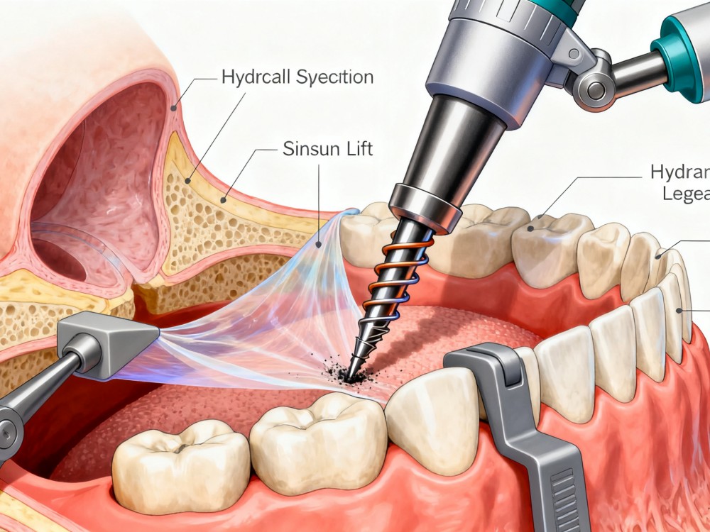
Special drill design ensures safer hydraulic membrane elevation during osteotomy procedures.
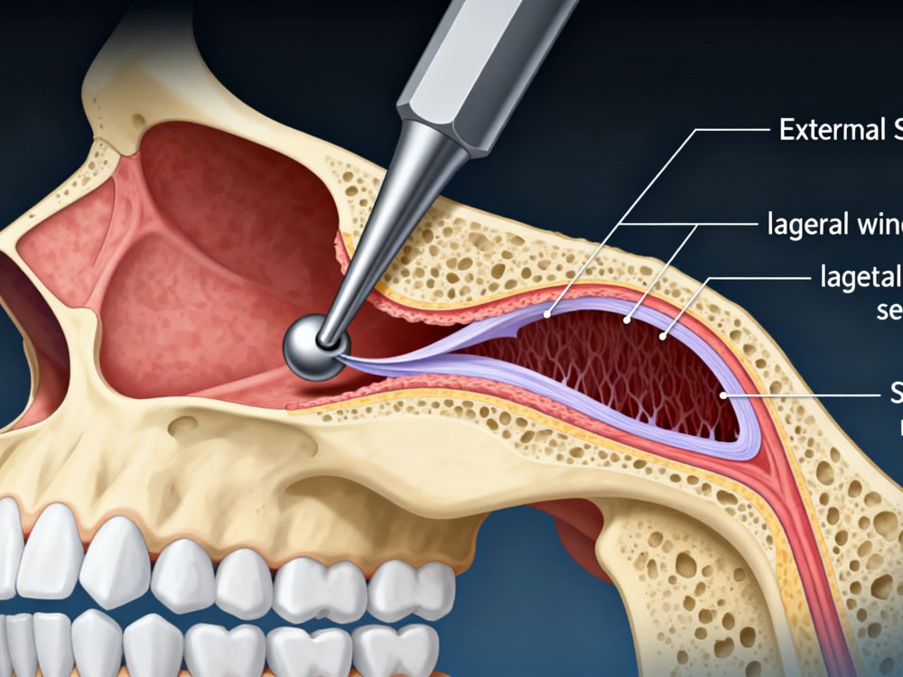
By opening a lateral window in the sinus wall, the use of sinus lifters allows safe membrane separation.
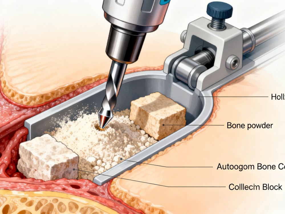
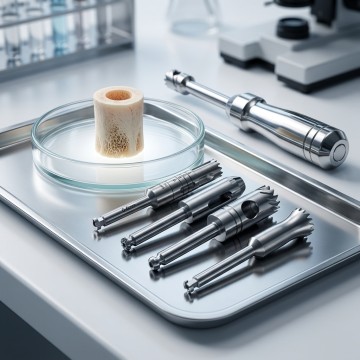
The hollow drill design minimizes bone loss and enables the collection of both autogenous bone powder and bone blocks.
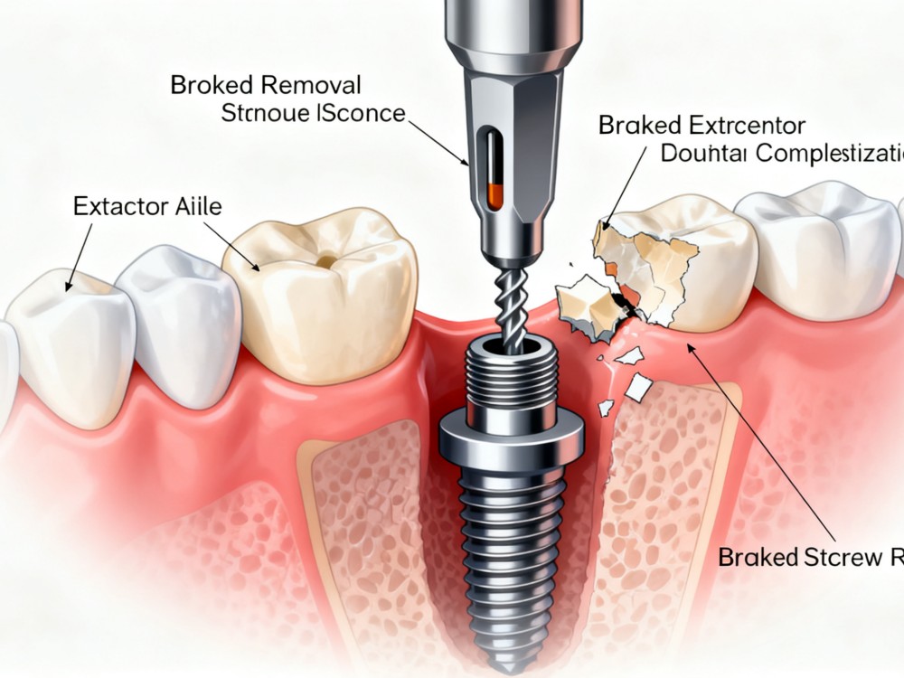
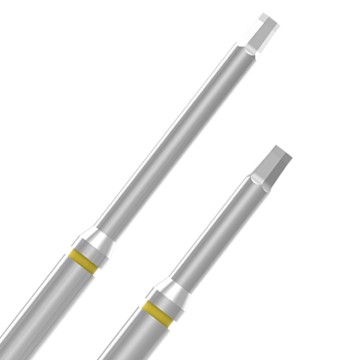
Whether it is a fractured abutment screw, central screw, or stripped screw, we provide comprehensive removal solutions.
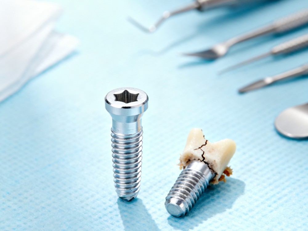
We offer solutions for extracting both fractured/damaged implants and intact implants.
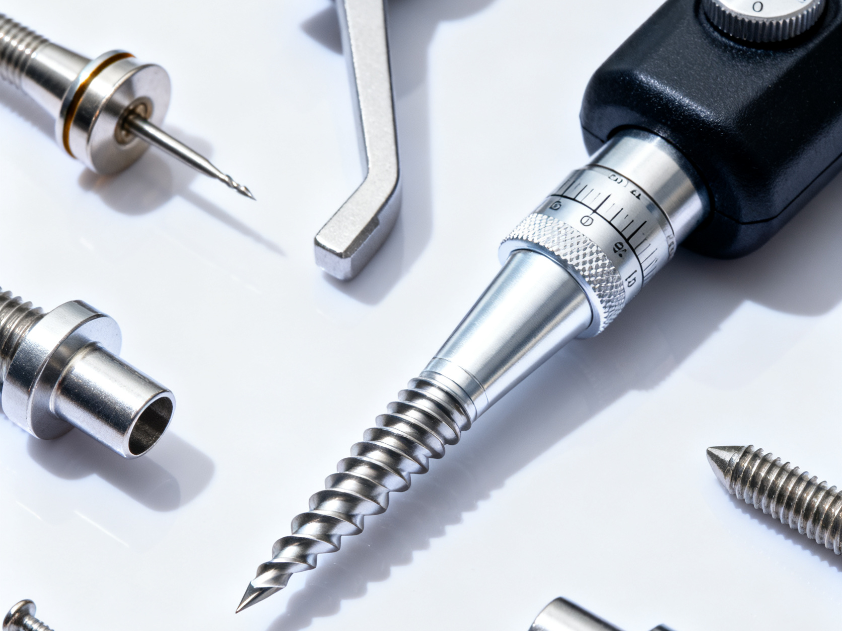
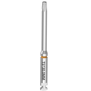
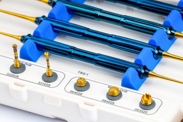
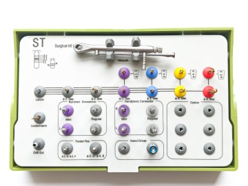
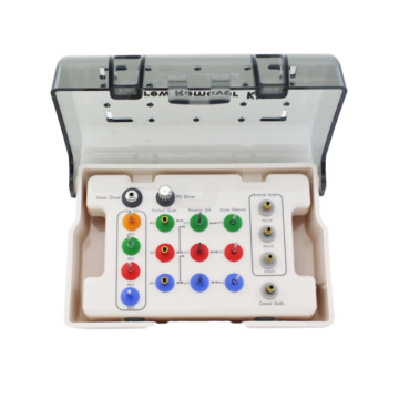
Our product range includes implant drills, torque wrenches, and related accessories to support the placement of different implant systems.
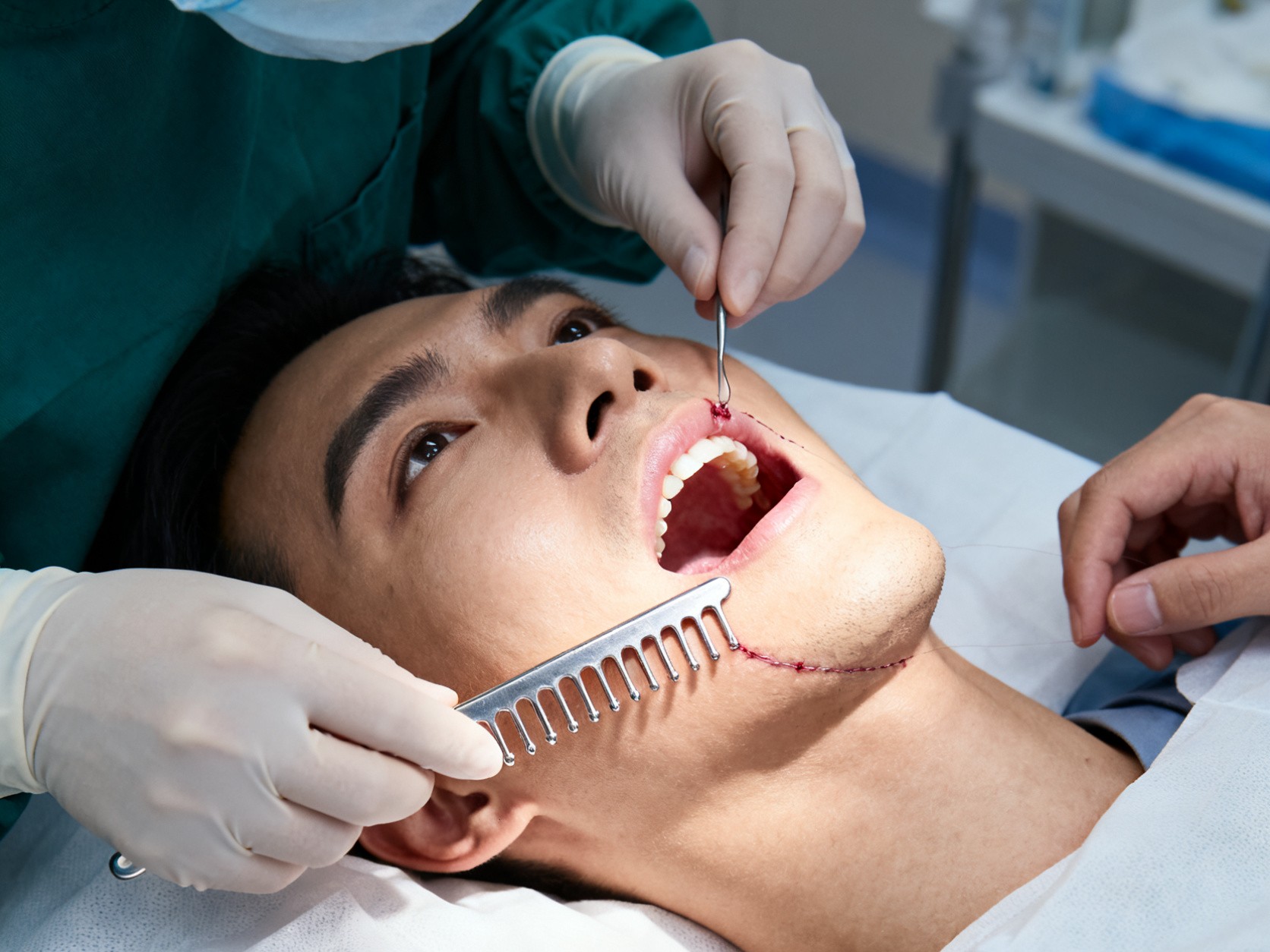
Our tension comb is designed for gingival soft tissue extension procedures.
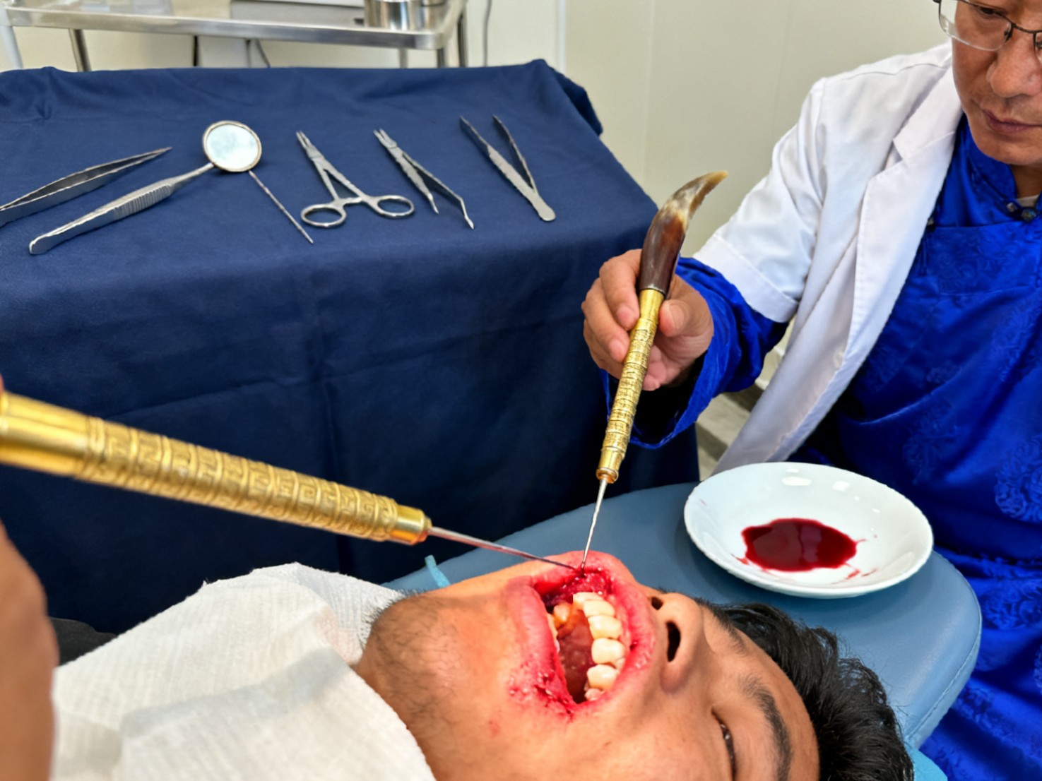
Our triangular needles are also applied in acupuncture and bloodletting therapy.
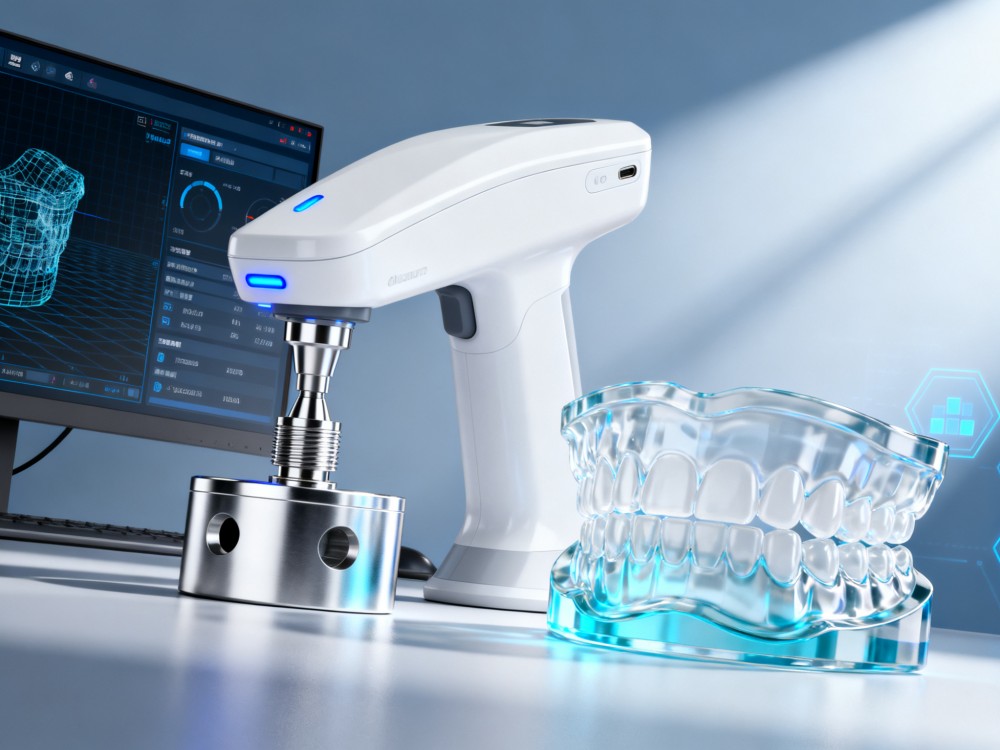
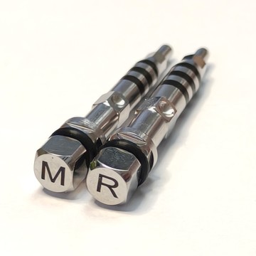
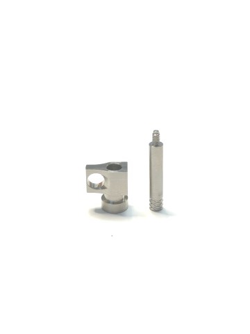
We provide customized scan bodies, scan abutments, and 3D analogs, integrated with databases to support your digital workflow.
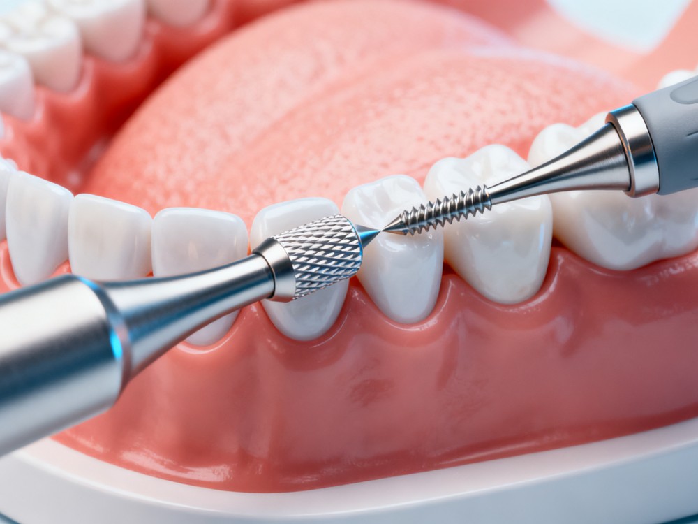
In combination with bone expansion drills, our tools facilitate shaping and controlled expansion of the alveolar ridge.
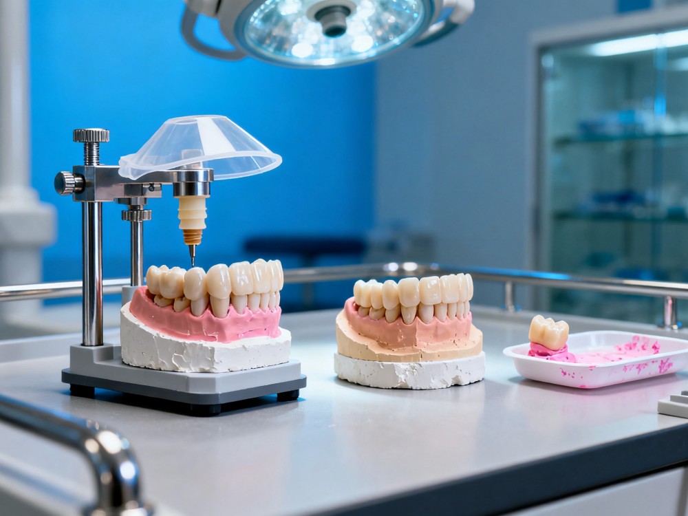
Our analogs, transfer posts, temporary abutments, and protective caps ensure comprehensive support.

Our instruments assist students in simulated surgery practice, clinical teaching, and hands-on training.
Strict quality control processes ensure that all digital components and surgical tools meet international standards.
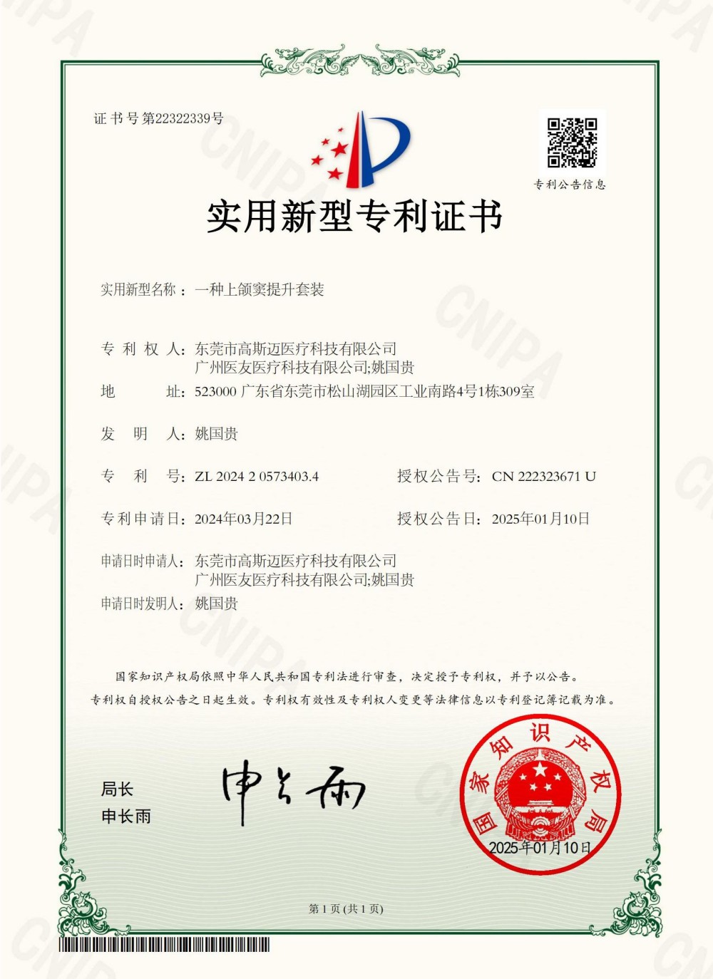
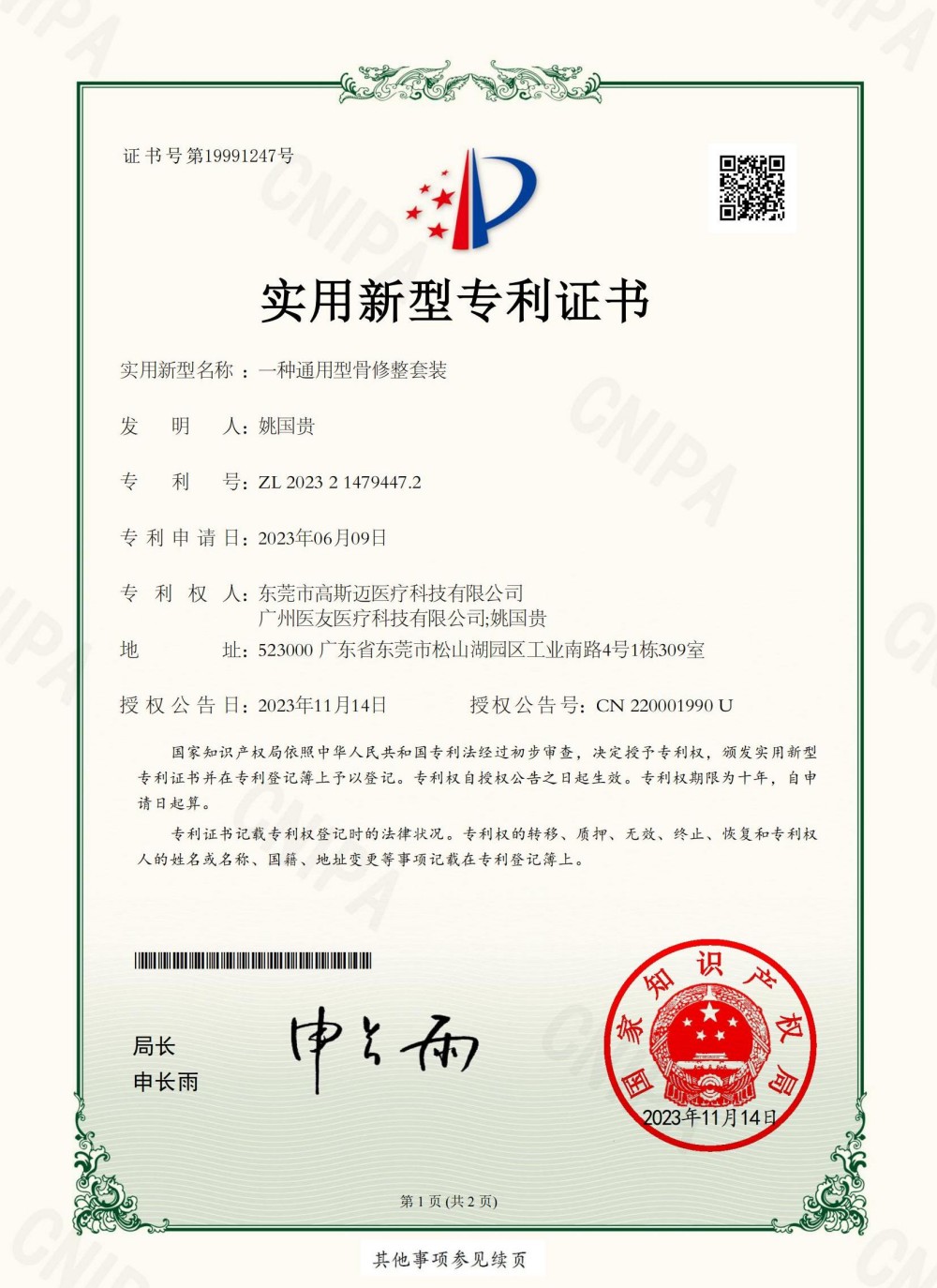
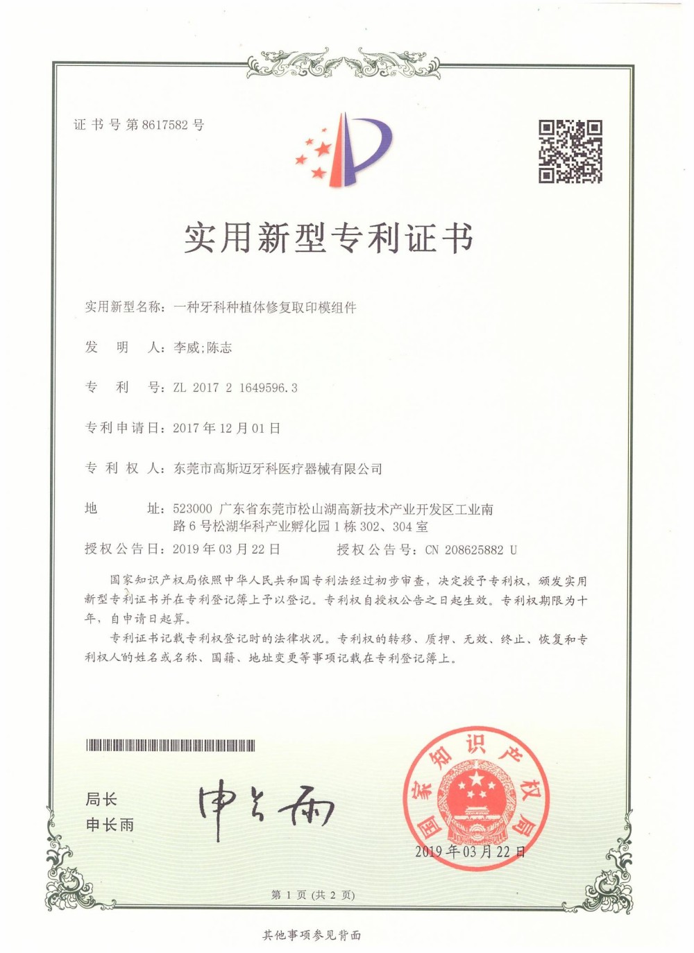
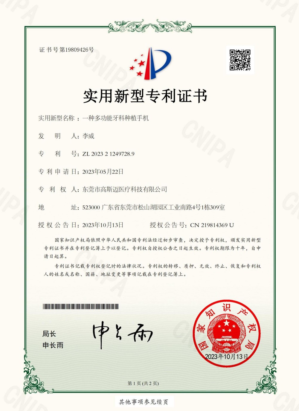
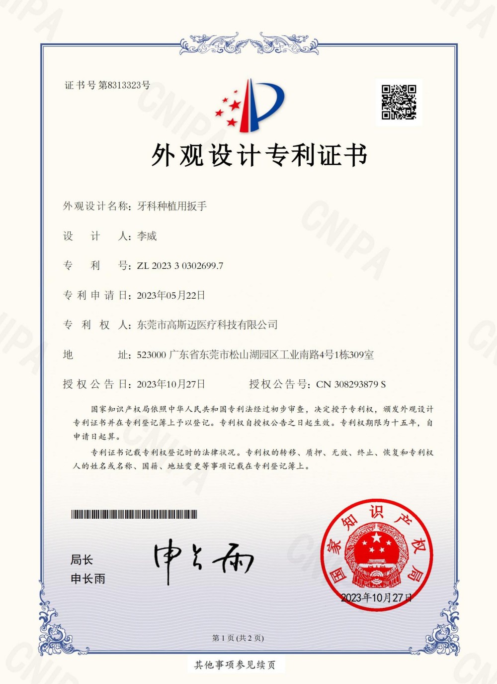
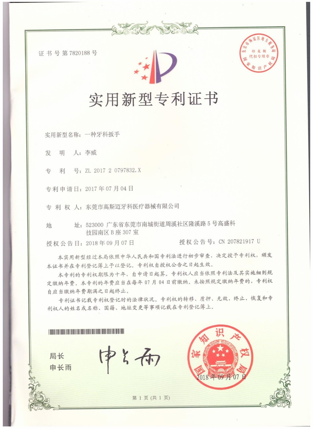
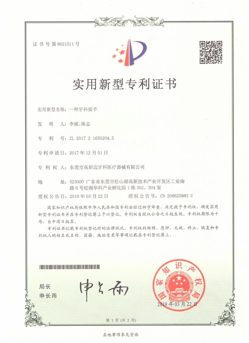
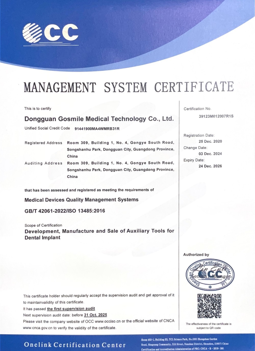
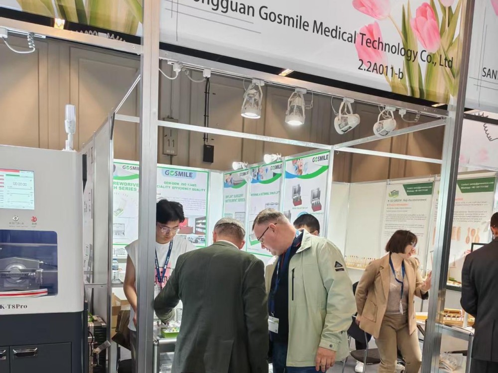
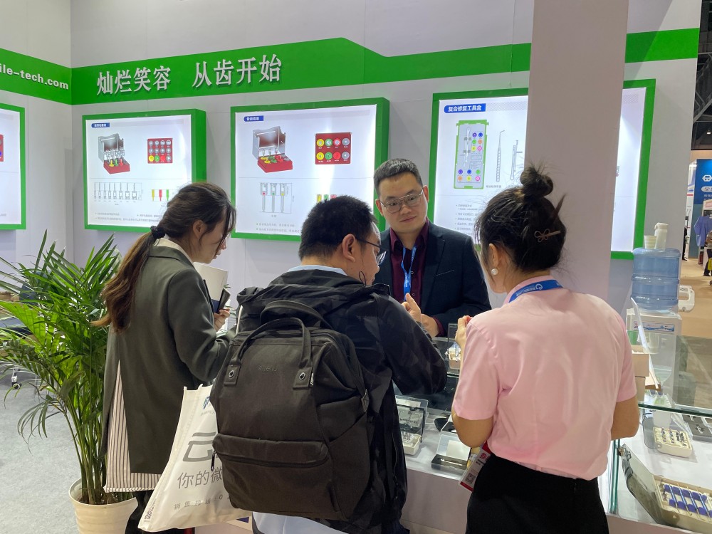
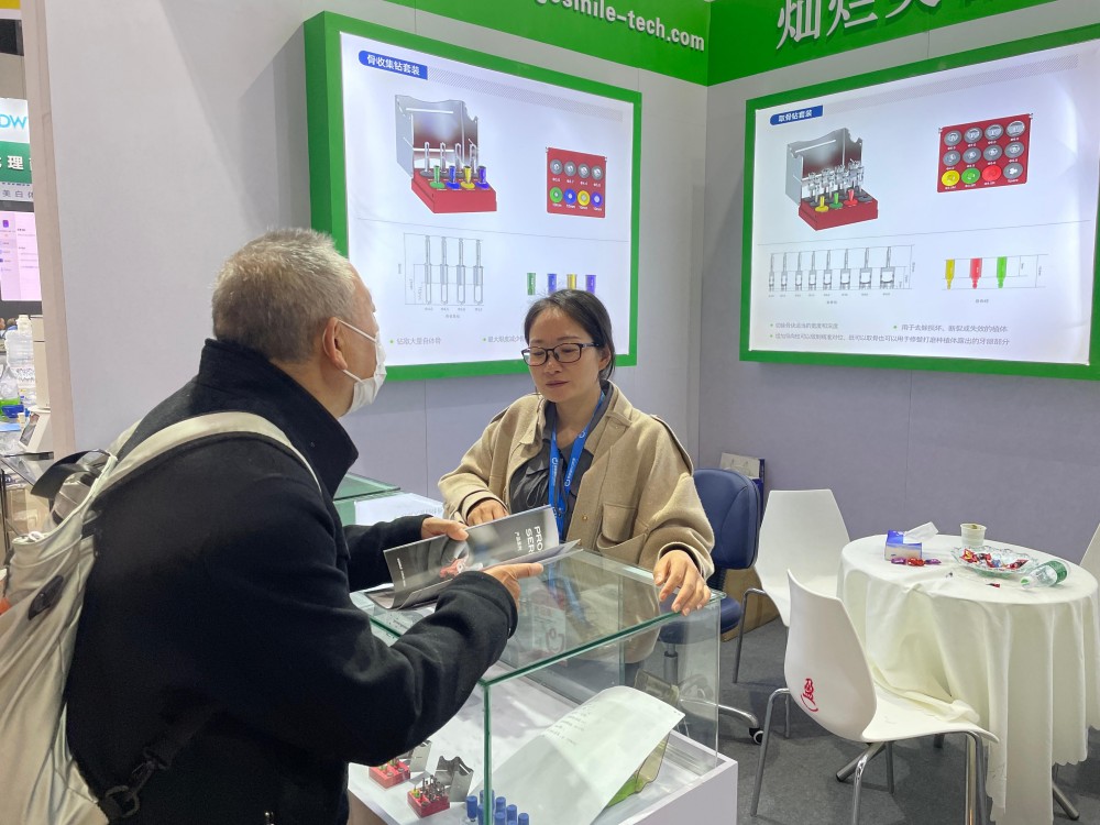
Connecting with dental professionals worldwide to showcase our latest innovations in digital implantology.


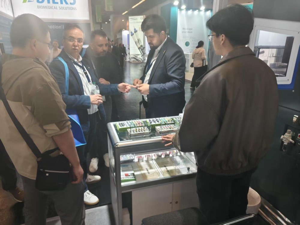
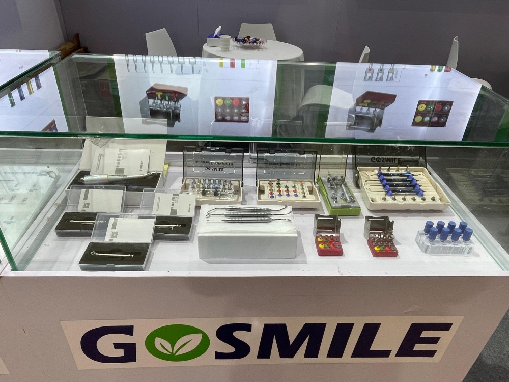
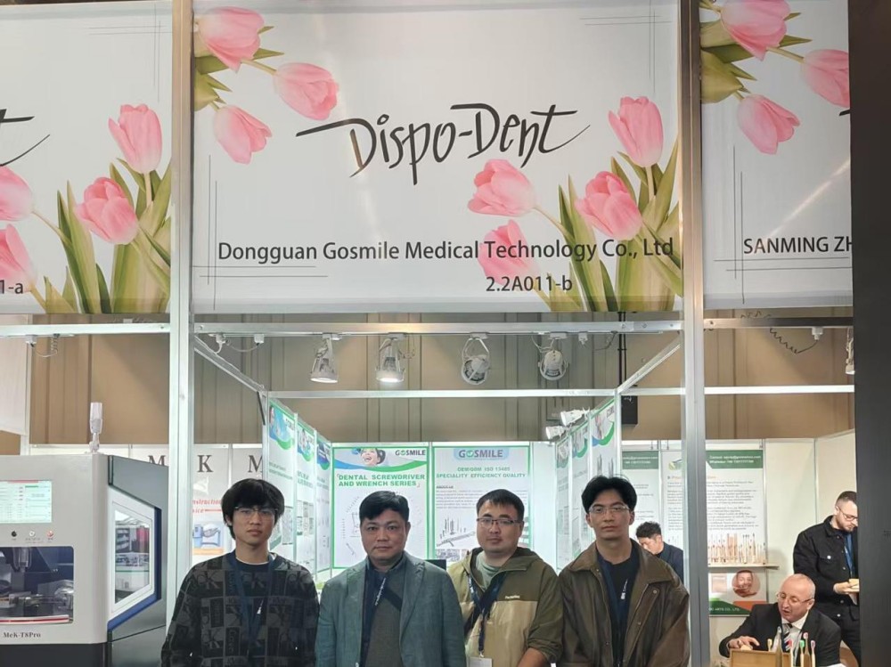

Explore our full range of high-precision drivers, kits, and scan accessories designed for implant placement and osteotomy.