Leave Your Message
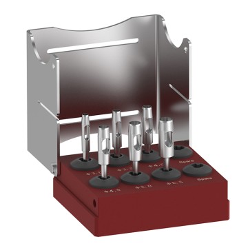
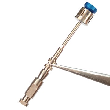
High-performance calibrated torque controls, depth-locked parallel pins, and ergonomic surgical drivers tailored for demanding trans-cranial portals and digital scan body registration assemblies.
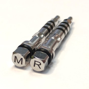
The modern paradigm of image-guided surgery (IGS) has traditionally relied on cumbersome optical tracking frames, invasive Mayfield cranial stabilization clamps, and skin-adhesive fiducial arrays. While effective for gross intracranial resections, traditional tracking mechanisms present intrinsic registration vulnerabilities during micro-craniotomy, skull base decompression, and keyhole cranial drilling. The historical boundary separating oral-maxillofacial implantology from neurosurgical access is dissolving. The cross-disciplinary implementation of high-precision dental scan bodies within cranial drilling pathways represents one of the most vital technological migrations in modern digital stereotaxy.
Originally engineered to digitize the exact spatial coordinate, angulation, and rotational orientation of osseointegrated dental fixtures, medical-grade dental scan bodies offer sub-10-micron dimensional repeatability. In cranial applications—ranging from temporal bone retrosigmoid approaches to anterior cranial fossa trephination and pterional craniotomies—these rigid titanium and polyetheretherketone (PEEK) fiducial geometries act as stationary spatial benchmarks directly fixed to rigid cranial landmarks. By securing custom scan body architectures into cortical bone burr margins or anchoring them via pre-planned micro-fixation plates, neurosurgical navigation suites achieve ultra-stable, non-shifting spatial registration that overcomes brain-shift error, scalp slippage, and optical line-of-sight obstruction.
Eliminates dynamic tracking drift caused by soft tissue mobilization. Rigidly anchored scan bodies preserve absolute mathematical coordinates between preoperative CT/MRI datasets and actual intraoperative cranial drilling paths.
CNC-machined from Grade 5 Eli Titanium with geometric tolerances within ±0.005mm. Compatible with intraoral scanners, optical desktop digitizers, and surgical infrared stereocameras.
Integration with mechanical stop keys, depth-guided sleeve bushings, and specialized trephine drills prevents mechanical plunging, safeguarding underlying dura mater, dural sinuses, and neurovascular bundles.
In cranial surgical access, the accuracy of automated navigation depends fundamentally on the rigid body transformation equation: P_patient = R * P_image + T, where R is a 3x3 orthonormal rotation matrix and T is a 3D translation vector. Standard adhesive fiducials yield registration errors ranging from 1.5mm to 3.5mm due to scalp elasticity, surgical prep fluid displacement, and positional shifts during patient drape positioning. In contrast, dental scan bodies affixed to anatomical hard tissue (such as zygomatic arches, supraorbital rims, or cranial anchoring micro-screws) provide non-yielding, geometrically unequivocal reference points.
The scan body features asymmetric chamfers, prismatic planar facets, and calibrated laser-etched barcodes that can be detected through optical intraoral scanners (IOS) or optical navigational cameras. The registration software aligns the known CAD library geometry of the scan body with intraoperative point-cloud scans using iterative closest point (ICP) algorithms. This workflow reduces Target Registration Error (TRE) to less than 0.45mm across the cranial operative field—an accuracy threshold critical for endoscope-assisted pituitary adenoma excision, microvascular decompression, and depth-restricted acoustic neuroma drilling.
1. Retrosigmoid & Far-Lateral Keyhole Approaches: In cerebellopontine angle (CPA) pathologies, drilling through the dense petrous bone requires razor-sharp trajectory verification. By securing a specialized dental scan body fixture onto the mastoid cortex, the surgeon establishes an instantaneous local reference system. The handpiece can be tracked dynamically, ensuring that burr-hole osteotomies avoid sinus laceration (transverse-sigmoid junction) while optimizing the viewing angle into the internal acoustic meatus.
2. Trans-Sphenoidal and Fronto-Orbital Access Guides: Complex craniomaxillofacial reconstruction often demands simultaneous intraoral and transcranial access. Using dental scan bodies intraorally on stable maxillary arches alongside temporary supraorbital scan body pins allows seamless multi-portal tracking. A singular navigational framework registers both the intracranial drill vector and maxillo-mandibular realignment plates, bypassing the need for separate coordinate references.
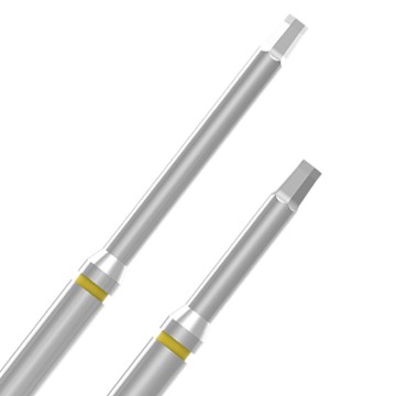
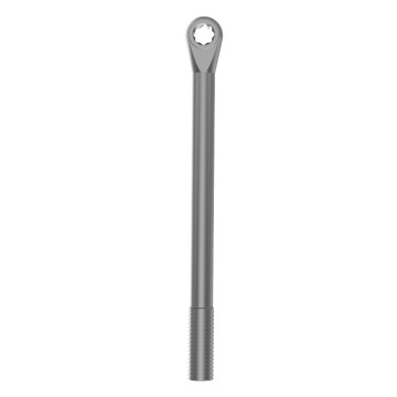
3. Stereotactic Depth-Controlled Fenestration in Cranioplasty: In revision cranioplasty and cranial trauma repair, drilling recipient bone margins for micro-titanium screw fixations risks penetrating fragile dural adhesions or scarred cerebral tissue. Incorporating torque-limited hex drivers, parallel guide pins, and scan-body-calibrated drill sleeves ensures the drill bit stops instantly at the exact prescribed cortical depth, safeguarding brain parenchyma while achieving optimal torque resistance.
From sterile CAD/CAM virtual simulation to high-precision 5-axis CNC micromachining and rigorous metrology validation, each surgical component meets stringent ISO 13485 medical standards.
 Consultation
 Development
 Production
 Inspection
 Sample
 Packaging
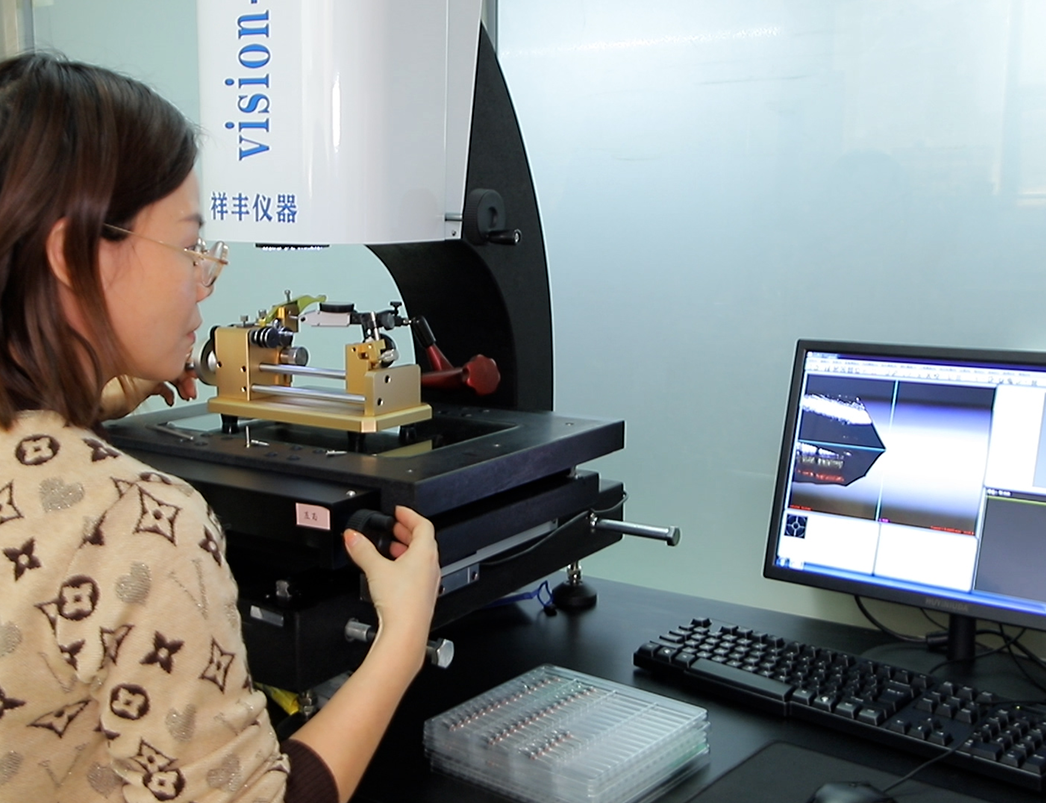
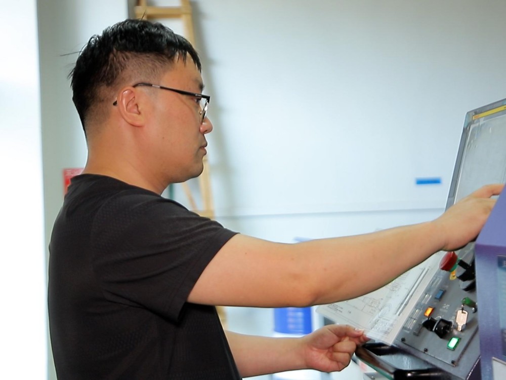
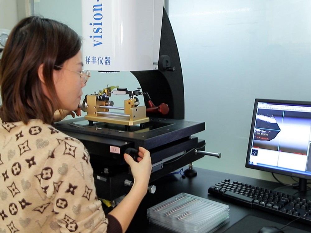
We implement a rigorous quality management system to ensure every batch undergoes thorough inspection and complies with standards. Utilizing advanced optical coordinate measurement machines (CMM), dynamic runout sensors, and high-vacuum material analyzers, our scan bodies, parallel pins, and torque drivers undergo 100% verification for pitch, angular fidelity, and surface biocompatibility before surgical delivery.
Engineered to interface seamlessly with major global implant libraries, cranial fixator platforms, and surgical navigation ecosystems including Adin, Anthogyr, Straumann, Osstem, and T-Plus.


Dongguan Gosmile Medical Technology Co., Ltd. located in Dongguan, Guangdong, is a high-tech enterprise that integrates development, testing, production, packaging, and sales. Established in 2017, we hold management system certificates and multiple national invention patents. We are a trusted supplier to major domestic distributors, known for our quality and reputation.
We are committed to efficiently producing high-quality medical products. Our mission is to provide reliable medical solutions and to build our own recognized brand in the advanced medical technology and surgical devices industry.
Choosing a name for a company is one of the most important and challenging tasks for any entrepreneur, as it is believed to bring good fortune. Faced with a myriad of options, our CEO adheres to a simple yet profound principle: simplicity is key. “Gosmile” is more than sufficient.
Once, I witnessed a touching scene on the street: a boy was looking at a girl with pure joy in his eyes. However, the girl, feeling insecure about her teeth, kept her hand over her mouth, afraid to smile. She worried that her smile wouldn't be beautiful enough. This moment inspired me to venture into the dental industry. I wanted to bring smiles to people like this girl, to help them gain the confidence to laugh without hesitation.
Today, that founding commitment to restoring natural facial harmony and physiological confidence drives our continuous R&D. Whether crafting ultra-precise scan bodies that navigate life-critical cranial osteotomies or manufacturing micro-fine dental implant drills, Gosmile engineers every product with unyielding compassion and aerospace-grade accuracy.
Leveraging digital manufacturing intelligence, global strategic alliances, and continuous surgeon feedback to advance medical device frontiers.
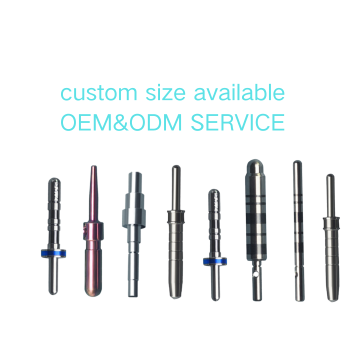
Data technology is inseparable from medical service. Based on market trends both domestically and internationally, as well as customer preferences, we offer customized products through OEM or ODM formats, leveraging data to create tailor-made solutions.
We collaborate with reputable suppliers and brand partners. These partnerships are crucial to our success. We provide them with high-quality products and services, and in return, they support and endorse our efforts.
We value customer feedback. We strive to uphold the positive evaluations from our clients, humbly accept any negative feedback, and commit ourselves to making continuous improvements.
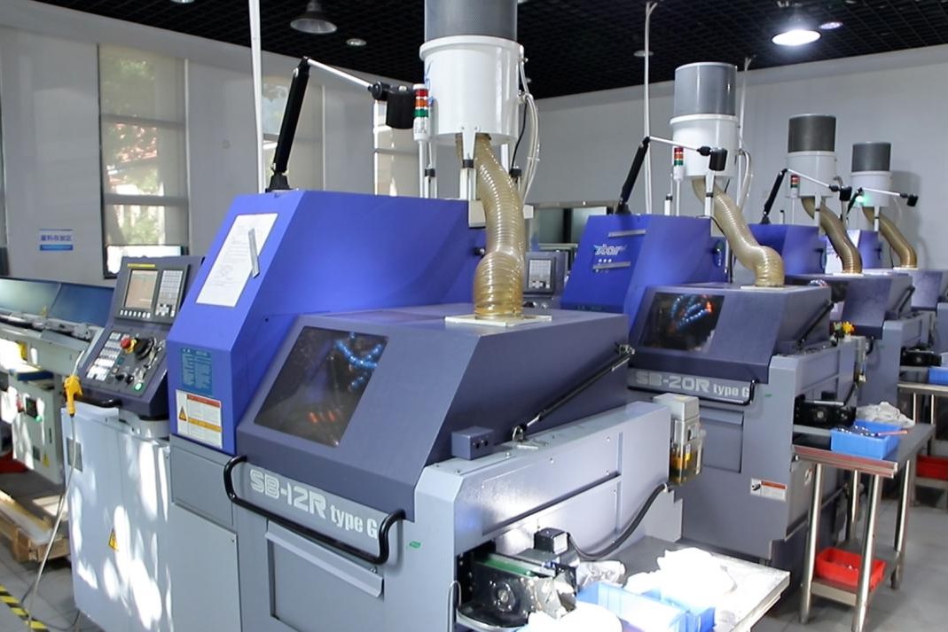
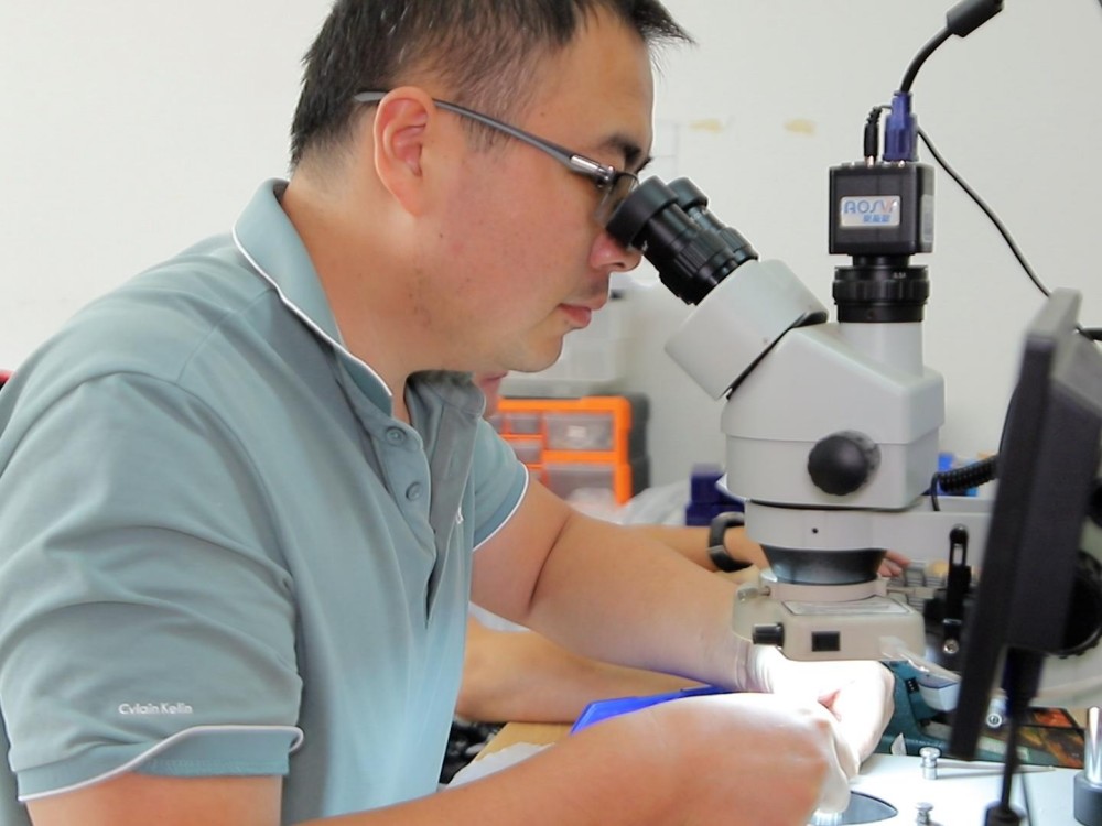
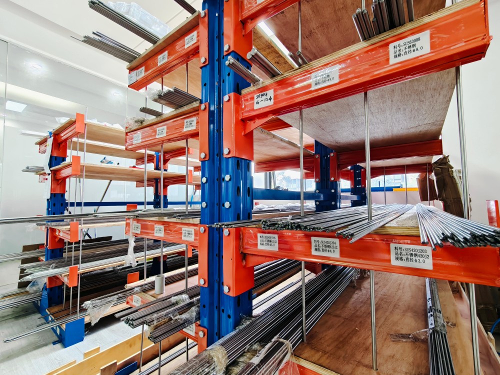
Our end-to-end production ecosystem guarantees full traceability, medical cleanliness, and micron-level conformance across nine synchronized milestones.




The global computer-assisted surgical navigation and skull-base drill guide market is projected to expand significantly over the coming decade, propelled by increasing demands for minimally invasive neurosurgical interventions. The interface of scan bodies within this domain will be defined by three critical evolutionary vectors:
1. Artificial Intelligence Automated Scan Body Recognition (AI-SBR): Current stereotactic cameras require high contrast and manual or semi-automated verification of scan body surfaces. Next-generation computer vision algorithms trained on thousands of synthetic volumetric datasets can instantly identify the spatial pose of fiducial scan bodies despite partial blood spatter, irregular illumination, or shallow angulations in deep surgical cavities. This drops registration latency to less than 100 milliseconds, allowing real-time re-registration should the operating table or navigation camera move.
2. Augmented Reality (AR) Overlay via Micro-Optical Scan Anchors: Wearable surgical head-mounted displays (HMDs) demand sub-millimeter tracking accuracy to project intracranial 3D structures (such as aneurysms, internal carotid arteries, and motor speech cortices) directly onto the operative field. By utilizing miniature titanium scan bodies as high-contrast anchoring beacons, AR glasses can maintain jitter-free holographic projection during active bone burring and osteotomy.
3. Regulatory Evolution & Biocompatible Nanomaterials: Cross-domain application requires components that comply with stringent dual-clearance medical device standards (such as FDA 510(k) Class II pathways and EU MDR Class IIb designations). Advances in radiopaque carbon-reinforced PEEK and ceramic zirconia scan bodies eliminate CT scattering artifacts, enabling flawless post-drilling intraoperative CT verification without image-degrading streaks.
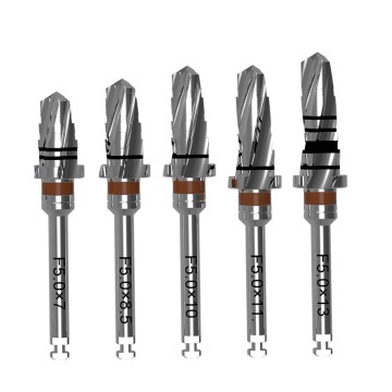
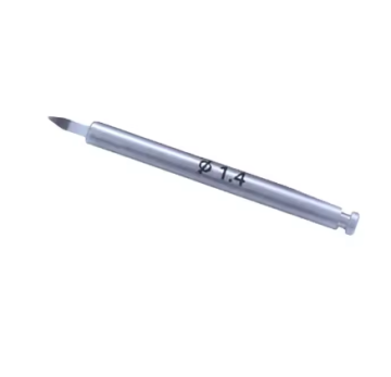
Explore our comprehensive inventory of machine-calibrated surgical drivers, restorative torque wrenches, bone-nourishing drills, laboratory analogs, and precision parallel pins.