Leave Your Message
Engineered to handle complex implant component failures with minimal bone loss and maximum clinical control.
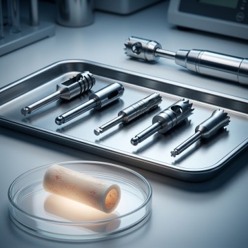
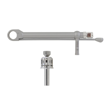
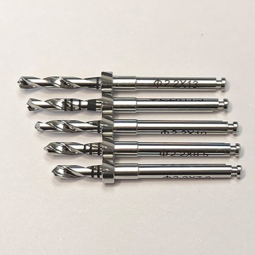
In the rapidly advancing landscape of restorative dentistry, dental implants have become the gold standard for replacing missing teeth. Despite high success rates exceeding 95%, mechanical complications inevitably arise. Among these, a fractured or broken implant screw represents one of the most frustrating and technically demanding challenges for dental clinicians. When an abutment screw shears off deep within the internal threads of the implant fixture, conventional retrieval methods often fail. This is where the specialized dental trephine bur for broken implant screw removal steps in as an indispensable rescue instrument.
Historically, a broken screw meant either leaving the fragment in place (rendering the implant useless) or executing a highly traumatic surgical block resection of the entire implant alongside surrounding bone. Today, advanced metallurgy and precision manufacturing have yielded specialized trephine burs designed to remove broken screws and osseointegrated fixtures while preserving maximum alveolar bone architecture. Understanding the engineering, clinical protocols, and market dynamics of these tools is critical for modern oral surgeons, periodontists, and implantologists.
Engineered with wall thicknesses under 0.5mm to minimize bone loss and target the implant-bone interface precisely.
Manufactured from high-strength surgical stainless steel or titanium alloys for superior cutting efficiency and durability.
Clear, high-contrast depth bands prevent accidental perforation of vital anatomical structures during surgery.
Implant screws are subjected to immense, repetitive masticatory forces within the oral cavity. Over time, these dynamic loads can lead to material fatigue. The primary causes of screw fracture include:
When a screw fractures, the top portion usually falls out, but the apical segment remains lodged inside the internal well of the implant. If the screw is broken below the level of the implant head, standard screwdrivers cannot engage it. In cases where the screw has cold-welded or stripped the internal threads, utilizing a dental trephine bur to isolate or extract the component (or the entire fixture if the internal wall is damaged beyond repair) becomes the definitive clinical solution.
When selecting a trephine bur for broken implant screw or fixture removal, always choose the smallest possible outer diameter that can clear the implant circumference. Preserving the buccal and lingual cortical plates ensures adequate bone volume for immediate or delayed replacement implants.
A dental trephine bur is a hollow, cylindrical drill bit featuring cutting teeth at its working tip. The design allows it to core around a cylindrical object—such as a broken screw, a post, or an entire implant fixture—without cutting into the core object itself. This makes it uniquely suited for atraumatic bone and metal drilling.
Traditional trephines used for bone grafting possess relatively thick walls. However, trephines optimized for implant removal feature ultra-thin walls. This minimizes the amount of bone removed at the periphery of the implant. The cutting teeth are typically designed with alternating bevels or diamond-impregnated surfaces to cut through both cortical bone and tough titanium alloys efficiently.
Matching the trephine to the implant system is critical. The internal diameter (ID) of the trephine must be slightly larger (typically by 0.1mm to 0.2mm) than the major diameter of the implant fixture or screw fragment. The external diameter (ED) must be kept as small as possible to limit the osteotomy size. High-quality manufacturers provide detailed compatibility charts matching their trephines to major implant systems like Nobel Biocare, Straumann, Zimmer, and ICX.
The global market for dental implant auxiliary tools is experiencing rapid growth, driven by the rising volume of dental implant placements worldwide. As the pool of patients with implants increases, the incidence of long-term mechanical complications, including screw fractures and implant failures, rises proportionally. This has transitioned the "implant rescue kit" from a niche luxury to a standard requirement for dental clinics.
From an industrial manufacturing perspective, the production of trephine burs demands extreme precision. CNC Swiss machining and advanced laser cutting are utilized to achieve the strict dimensional tolerances required for thin-walled trephines. Furthermore, the industry is witnessing a shift toward specialized surface coatings, such as Diamond-Like Carbon (DLC) or Titanium Nitride (TiN), which reduce friction, prevent overheating of the bone, and increase the lifespan of the cutting edge.
OEM and ODM manufacturing services have also expanded, allowing implant companies to include custom-branded trephine drills and extraction kits within their official surgical catalogs. This ensures that clinicians have access to tools specifically calibrated for their proprietary implant geometries.
When faced with a broken screw that cannot be retrieved using reverse-threaded screw extractors or ultrasonic tips, the clinician must proceed with systematic trephine extraction:
The future of implant rescue procedures lies in digital integration. Guided surgery templates, designed using pre-operative CBCT scans, are now being adapted for trephine applications. By utilizing a 3D-printed surgical guide, the clinician can lock the trephine bur into a precise trajectory, eliminating the risk of lateral deviation, which could damage adjacent teeth, nerves, or cortical plates.
Additionally, research into "smart materials" is paving the way for self-limiting drills that automatically stop cutting when they transition from hard metal to soft tissue or bone of different densities. These advancements promise to make the retrieval of broken implant components safer, faster, and more predictable than ever before.
Our dental implant auxiliary instruments and trephine drills support a comprehensive range of surgical interventions.
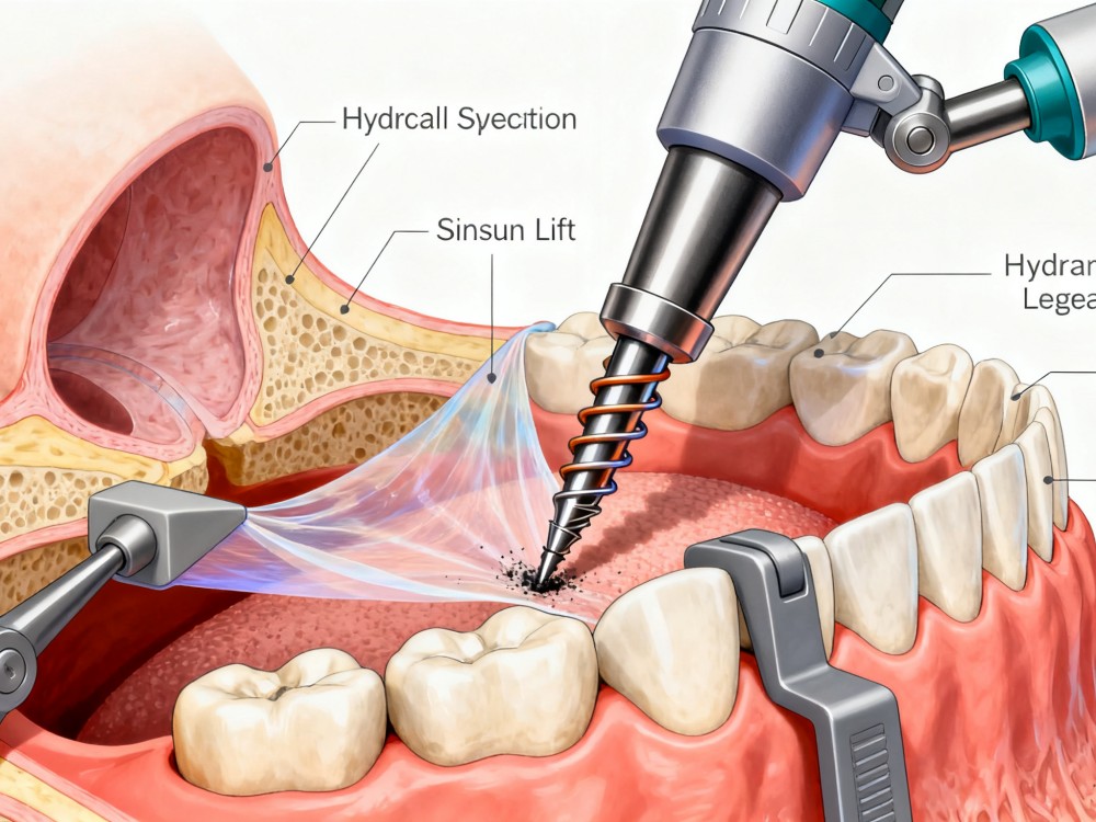
Special drill design ensures safer hydraulic membrane elevation during sinus floor elevation procedures.
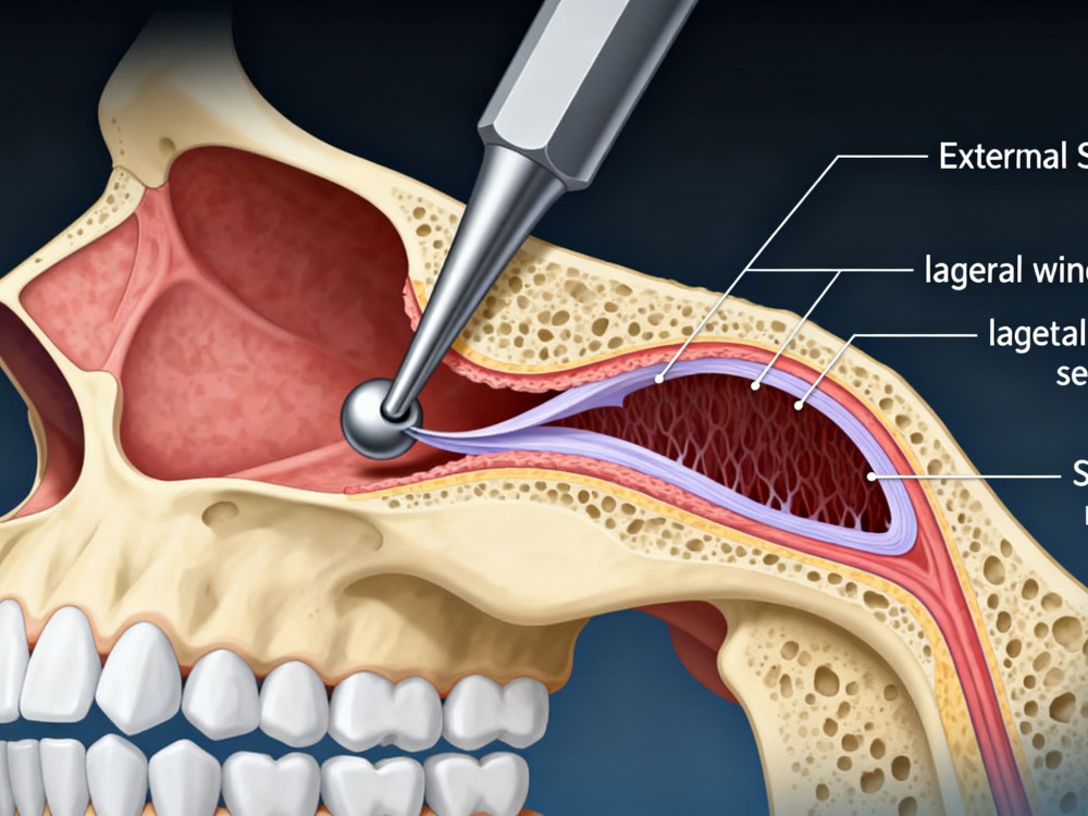
Safe membrane separation through a lateral window using advanced sinus lifters and specialized burs.
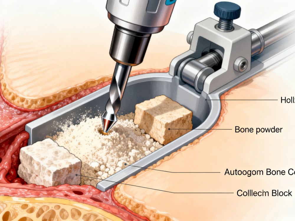
Hollow trephine design minimizes bone loss, collecting autogenous bone powder and bone blocks simultaneously.
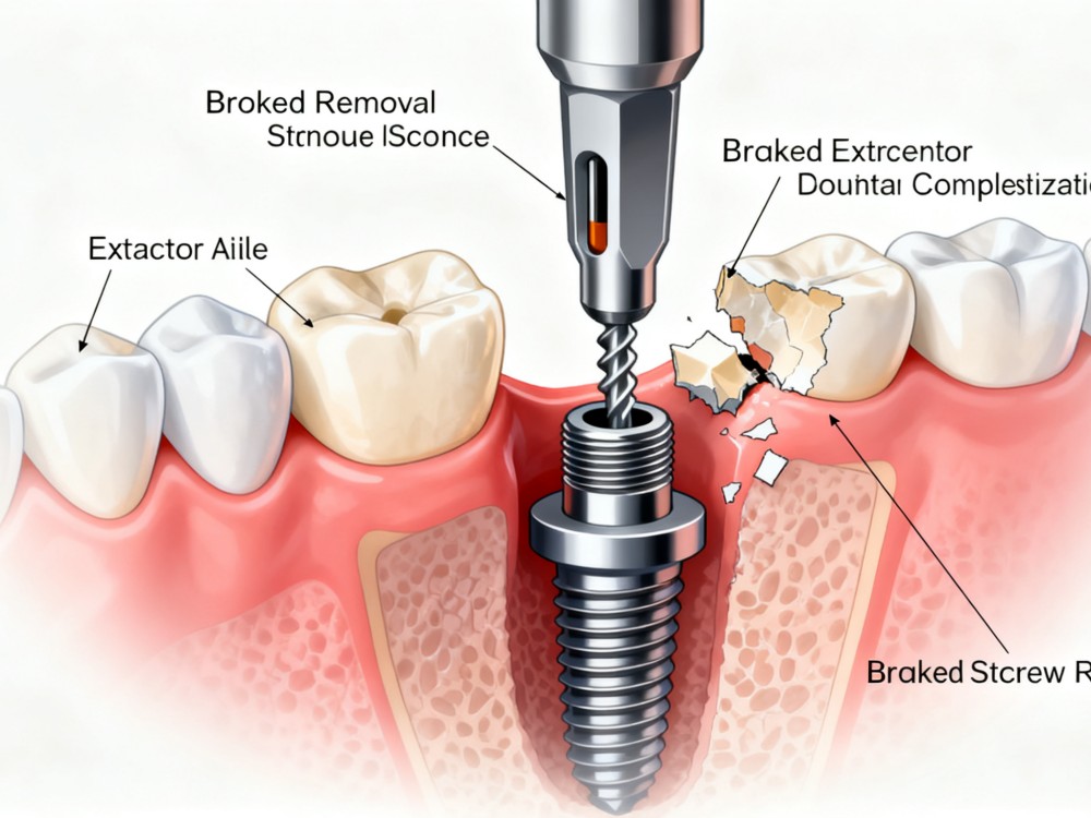
Comprehensive retrieval solutions for fractured abutment screws, central screws, or stripped prosthetic screws.
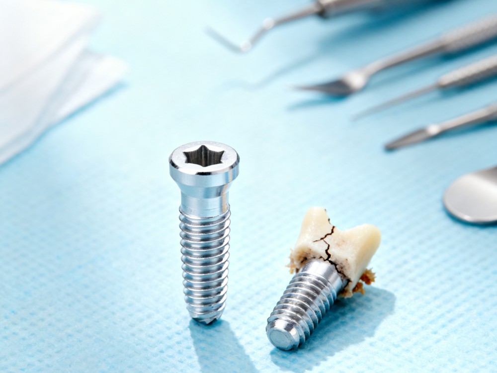
Atraumatic extraction solutions designed for both fractured, damaged, and fully osseointegrated intact implants.
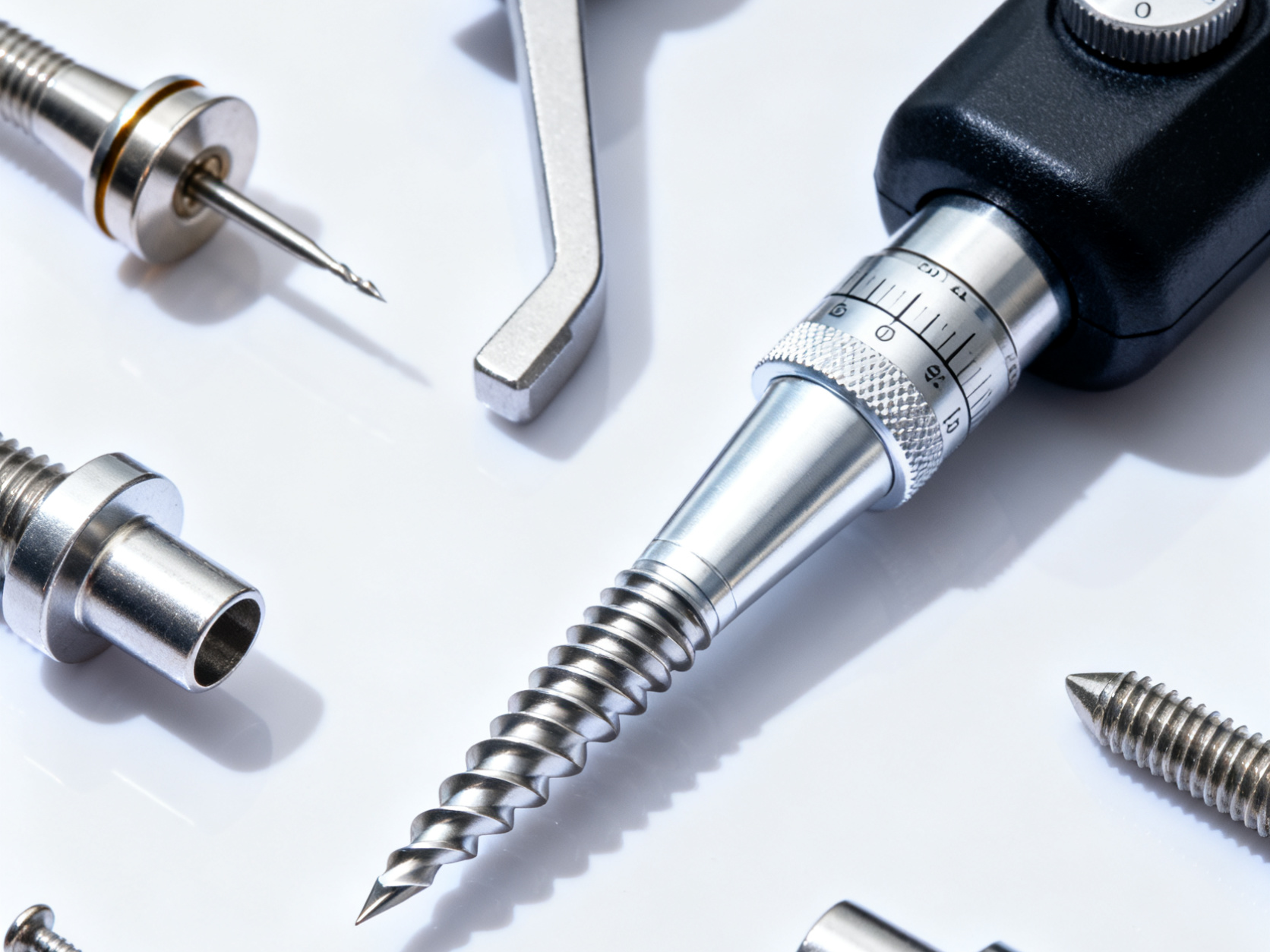
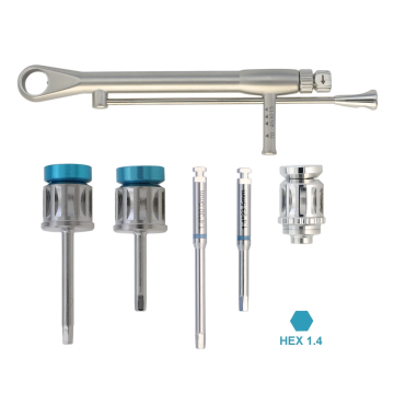
A complete line of drills, torque wrenches, and adapters supporting major global implant systems.
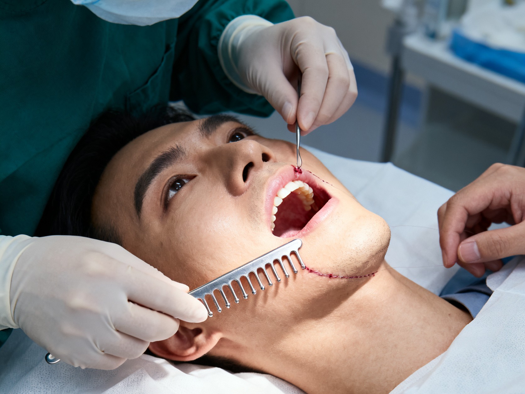
Innovative tension combs designed to safely extend gingival soft tissue during complex flap procedures.
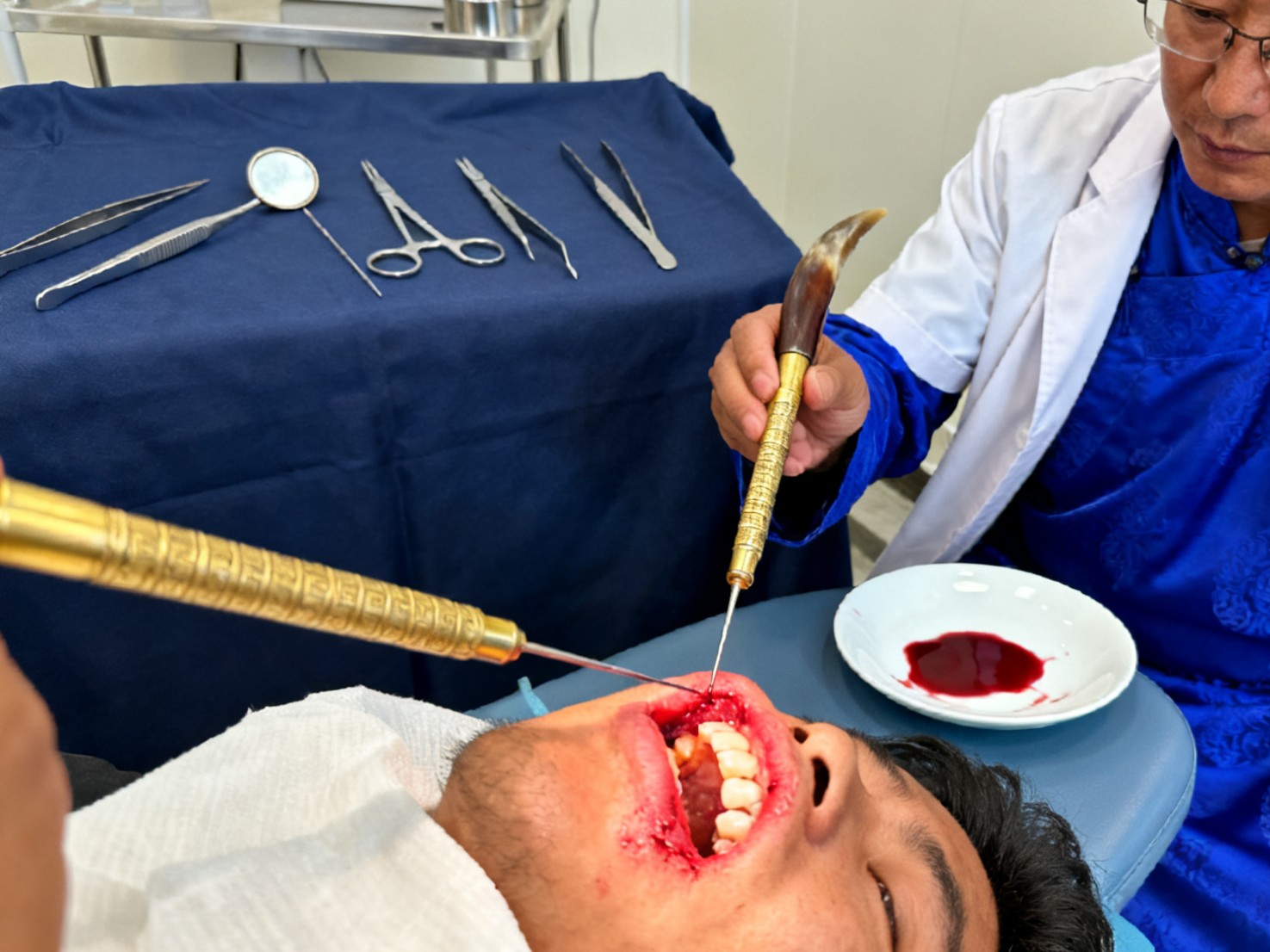
Precision triangular needles designed for acupuncture and traditional bloodletting therapy applications.
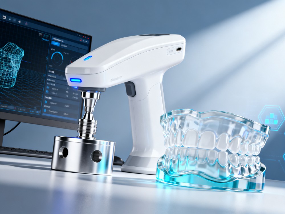
Customized scan bodies, scan abutments, and 3D analogs supporting integrated digital workflows.
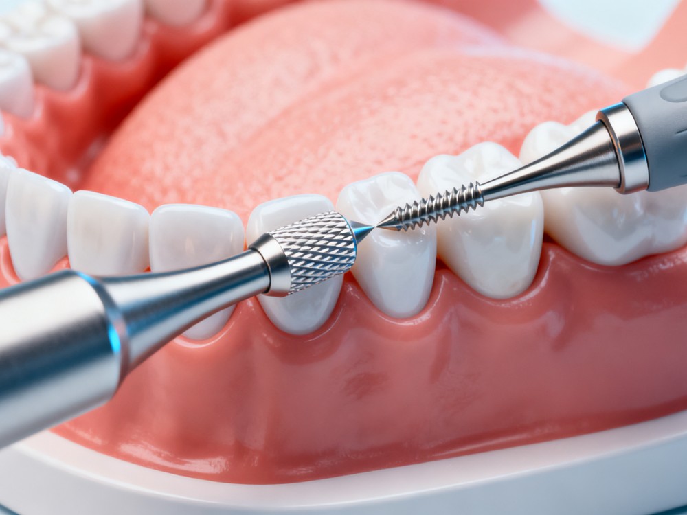
Controlled expansion and shaping of narrow alveolar ridges using advanced bone expansion drills.
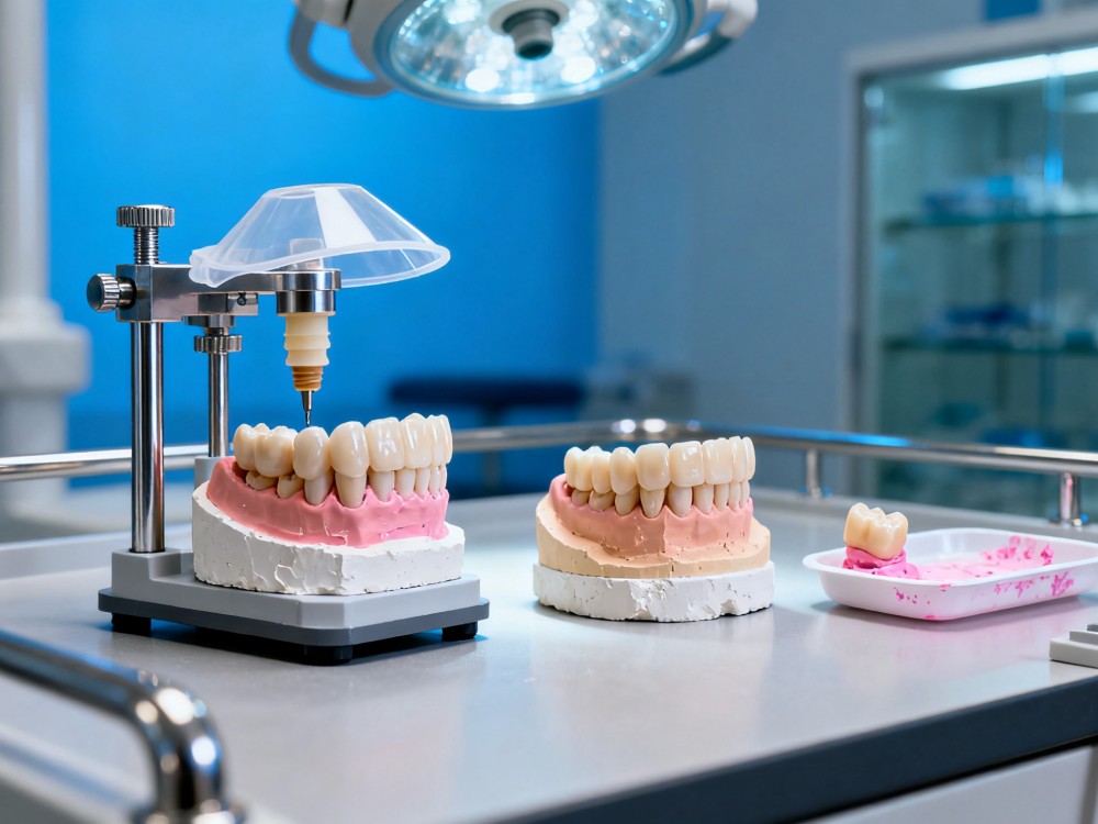
High-fidelity analogs, transfer posts, temporary abutments, and protective caps for restorative accuracy.

Simulated training models and instruments for university teaching, clinical hands-on seminars, and student practice.
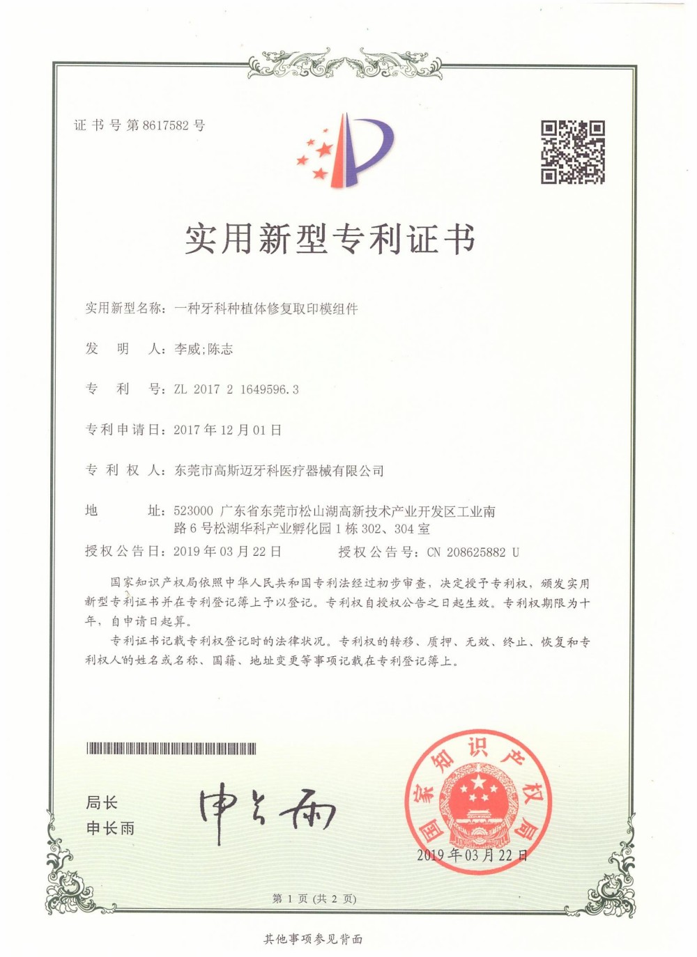
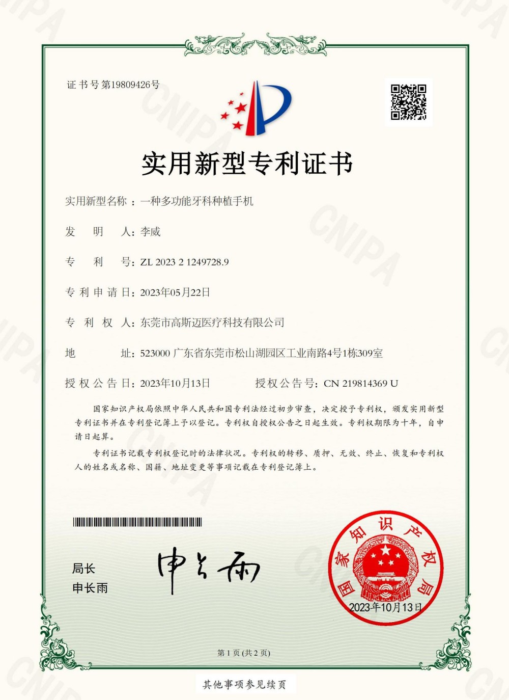
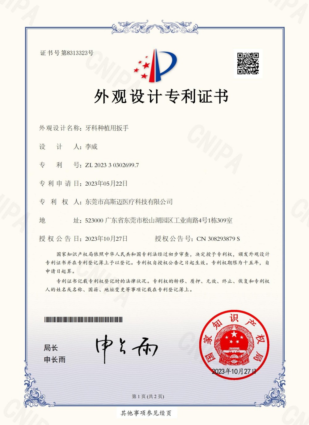
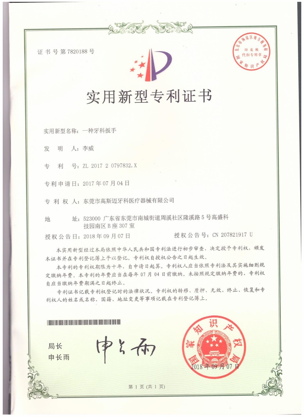
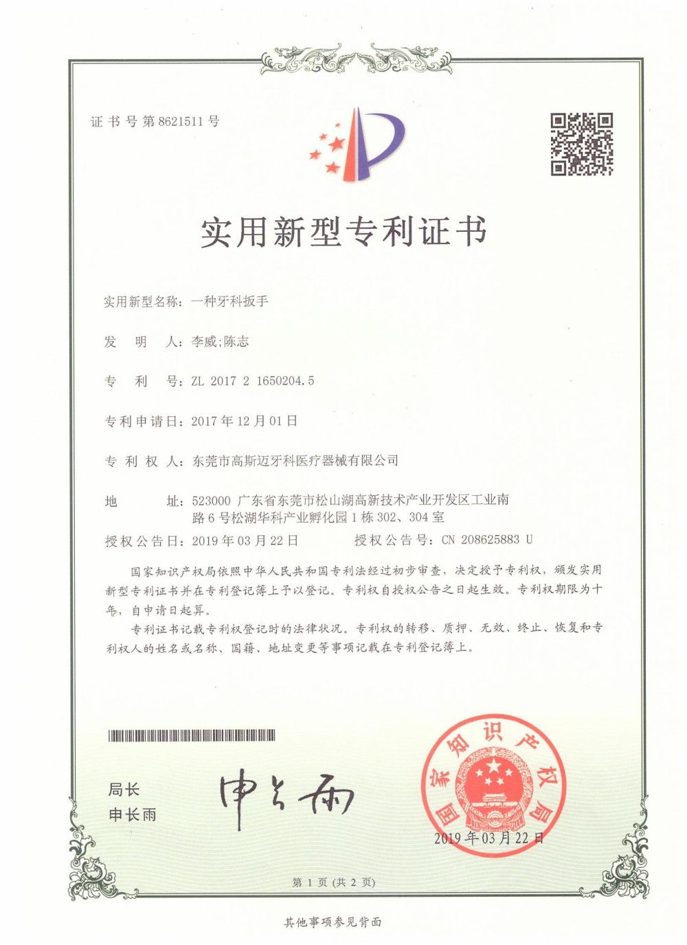
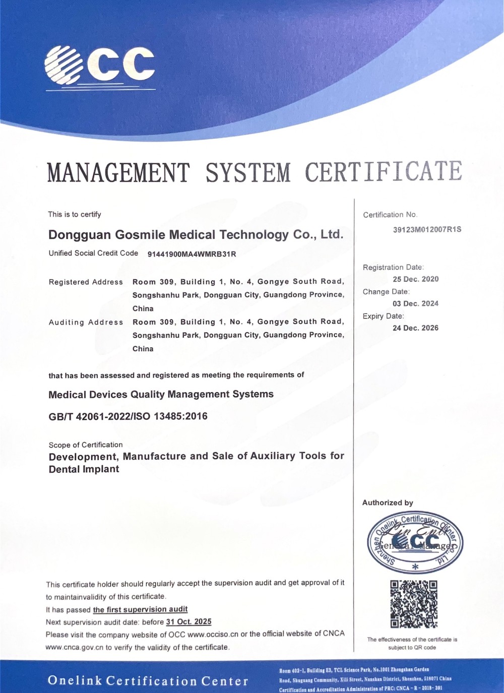
Our production line conforms to international medical standards to guarantee surgical safety and reliability.
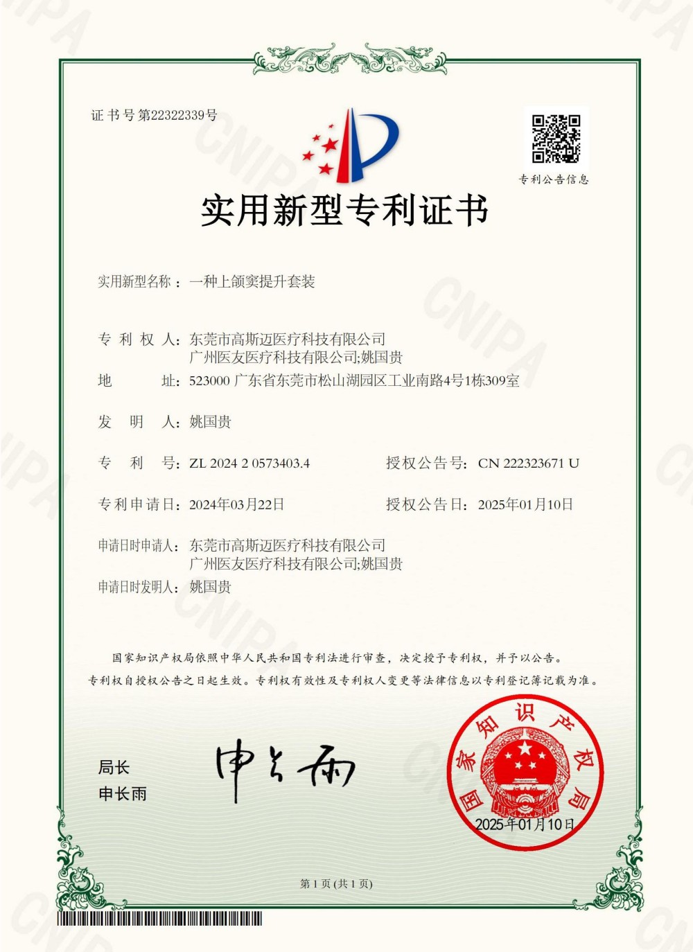
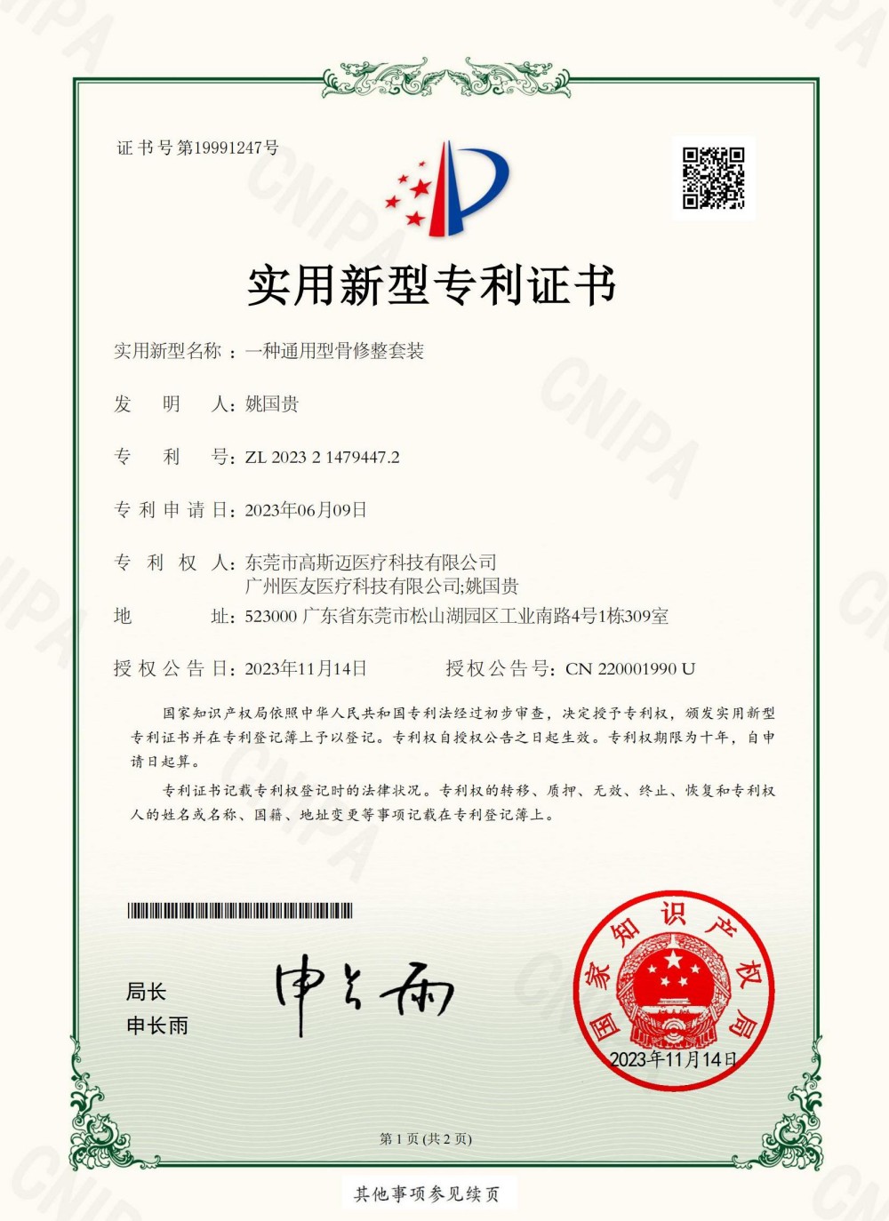
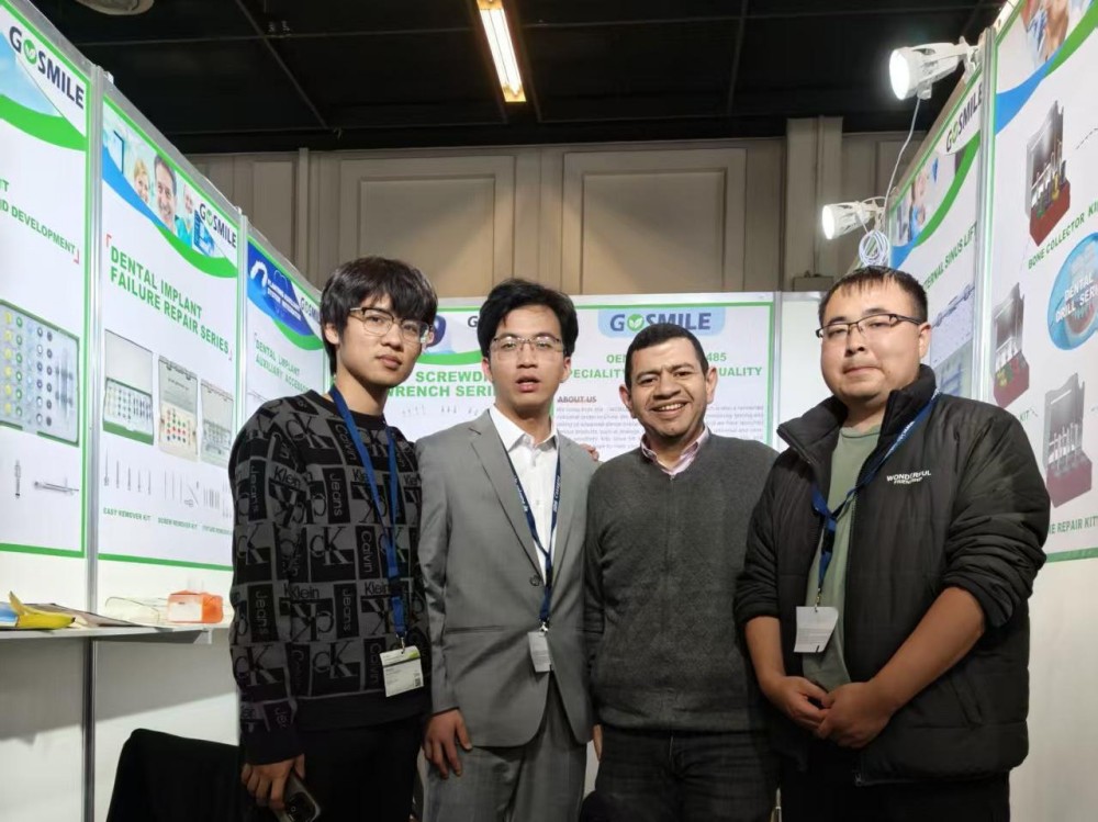
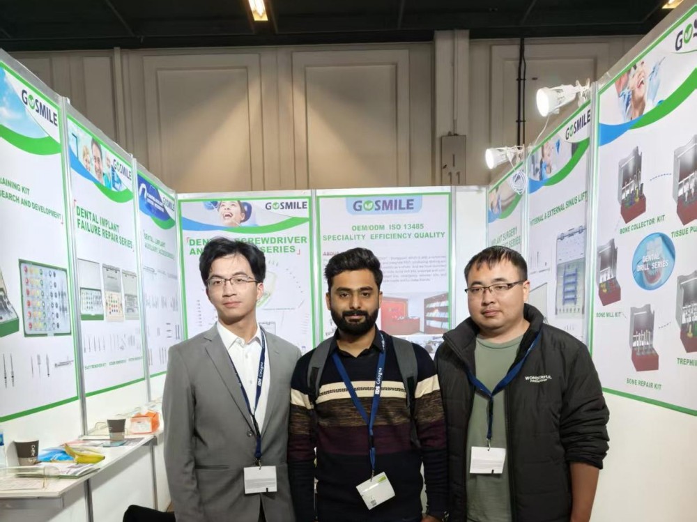
Connecting with dentists and medical distributors worldwide to demonstrate our latest implant solutions.
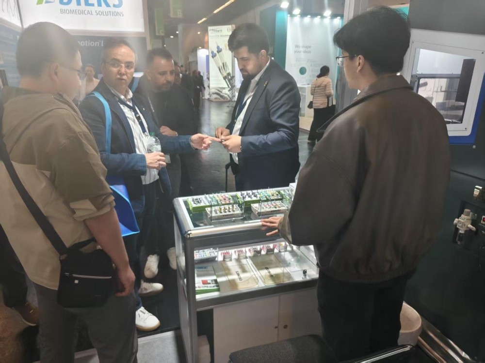
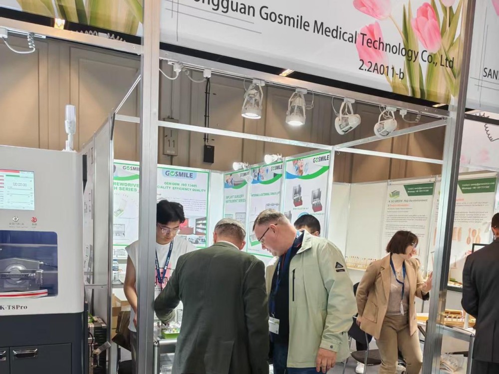
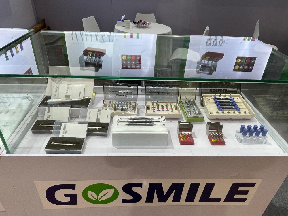
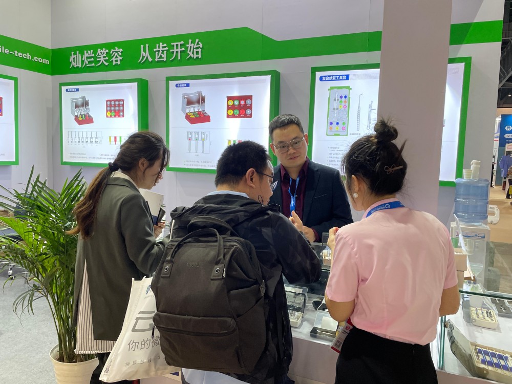
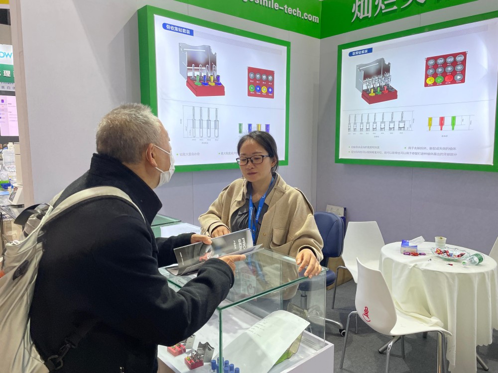
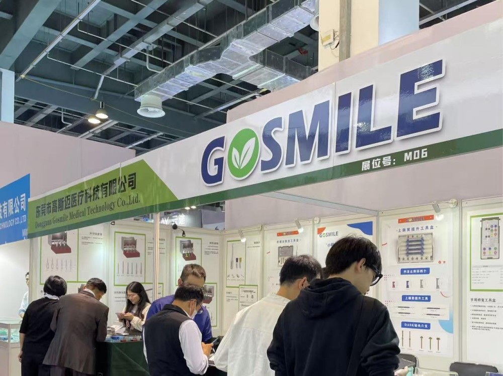
Explore our complete range of surgical drills, restorative drivers, and extraction instruments.