Leave Your Message
Modern implantology has evolved beyond simple tooth replacement. Today, clinicians routinely confront severe alveolar ridge resorption, requiring sophisticated bone augmentation procedures. Among these, autogenous bone harvesting and grafting remains the biological gold standard. However, the success of these complex surgeries relies heavily on the subsequent restorative phase. This is where the Dental Implant Analog plays a vital, yet frequently underappreciated, role in the CAD/CAM model workflow.
Unlike synthetic or xenogeneic materials, autogenous bone blocks harvested from intraoral sites (such as the mandibular ramus or symphysis) possess osteogenic, osteoinductive, and osteoconductive properties. This dual biologic activity accelerates graft consolidation, providing a robust foundation for implant placement.
When executing autogenous bone grafting, the spatial architecture of the site is completely altered. The newly grafted block or particulate bone must heal and integrate. Once integrated, the implant is placed. The role of the implant analog during the prosthetic fabrication is to replicate the exact three-dimensional position of the implant fixture within the laboratory model. Any minor deviation in the analog's seat can lead to mechanical stress on the newly grafted bone, potentially inducing bone resorption and implant failure.
The global dental implant market is experiencing a significant shift towards digital workflows. Historically, traditional plaster models utilized brass or stainless steel lab analogs. Today, the rise of 3D printing and CAD/CAM technologies has birthed a new class of digital analogs (often referred to as 3D model analogs or CAD/CAM analogs). These components are designed with tight tolerances to fit perfectly within 3D-printed resin models.
In the context of autogenous bone harvesting and grafting, the industrial demand for high-precision, anti-rotational implant analogs is higher than ever. Because grafted bone is more susceptible to micro-movements during the early loading phase, prostheses must fit with passive accuracy. Industrial manufacturers are now utilizing ultra-precise CNC milling machines and medical-grade Titanium Alloy (Grade 5) to produce analogs that match the implant-abutment interface with micron-level precision, minimizing prosthetic mismatch.
Consider a patient undergoing a block graft in the anterior maxilla due to severe traumatic bone loss. The surgical team harvests a cortical bone block from the ramus, secures it with fixation screws, and allows it to heal for 4 to 6 months. Following successful osteointegration of the graft, the implant is placed.
During the prosthetic phase, the clinician uses a digital scan body to capture the implant's position. In the laboratory, the digital file is imported into CAD software, where the technician designs the model and selects the corresponding digital implant analog from the library. The physical model is 3D printed, and the analog is inserted from the bottom of the model, secured by a precise locking screw. This physical-digital hybrid model allows the technician to build a custom abutment and crown that fits the patient's newly grafted tissue contour without creating tension. The precision of this analog directly determines the longevity of the autogenous bone graft, as uneven force distribution could lead to micro-fractures in the grafted bone matrix.
As artificial intelligence continues to integrate into dental software, we are seeing the emergence of smart libraries that automatically adjust analog parameters based on the density of the surrounding bone. In cases of autogenous bone grafting, where bone density might vary across the graft-host interface, CAD software can recommend specific analog insertion depths to optimize stress distribution. Furthermore, manufacturers are exploring color-coded anodization for titanium analogs to prevent cross-system errors in multi-platform laboratories, streamlining the workflow and reducing clinical margins of error.
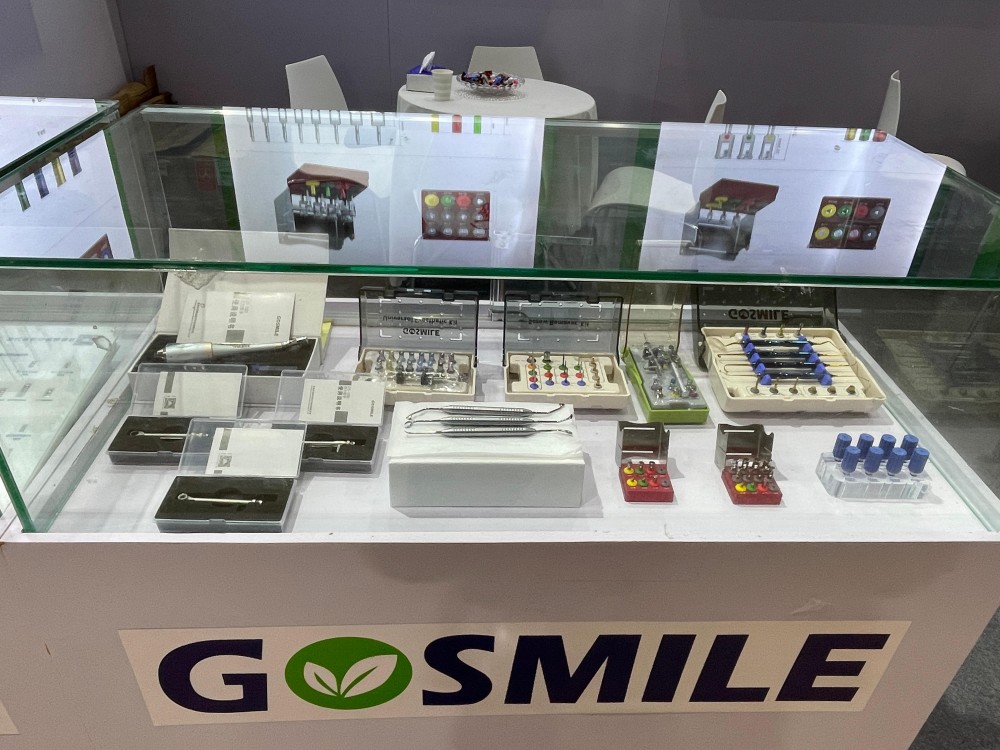
Our dental implant auxiliary tools and precision analogs are designed to support clinicians across a wide range of complex oral surgeries and laboratory workflows.
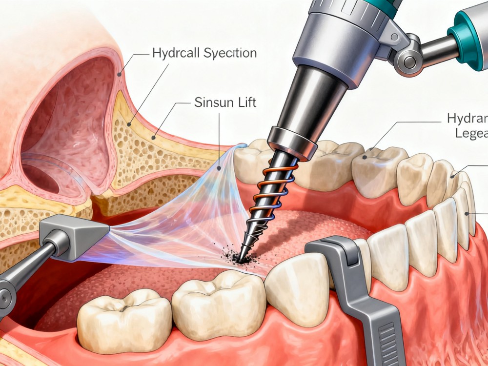
Special drill design ensures safer hydraulic membrane elevation, preparing the site for sinus grafting and subsequent implant analog planning.
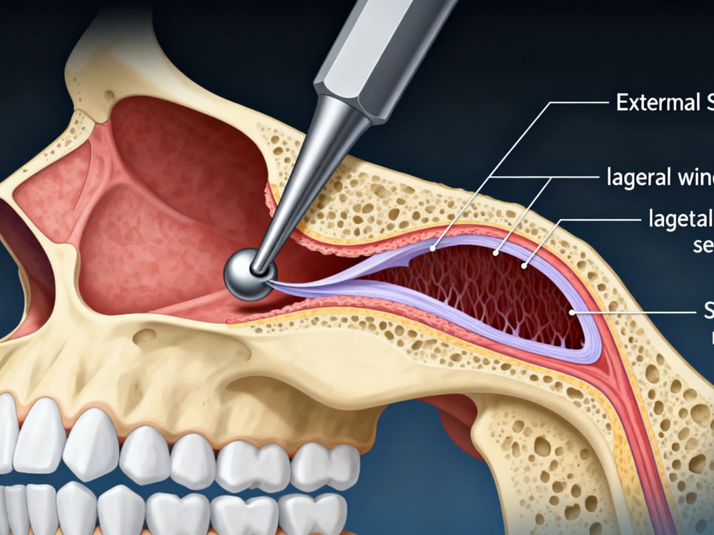
By opening a lateral window in the sinus wall, our sinus lifters allow safe membrane separation to pack bone graft materials effectively.
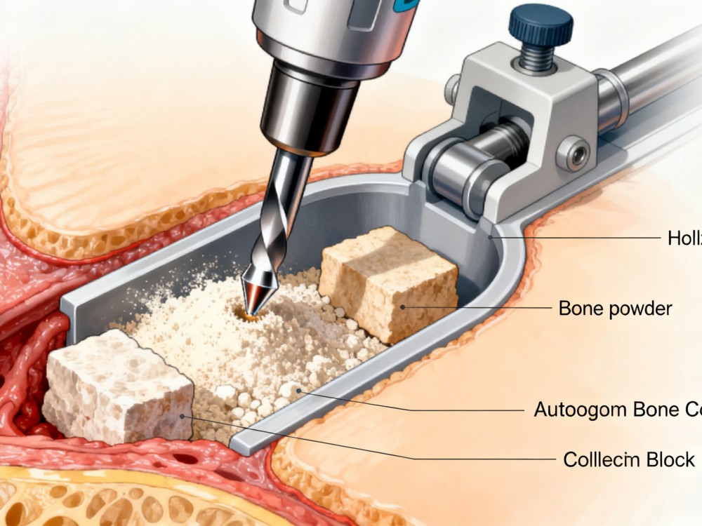
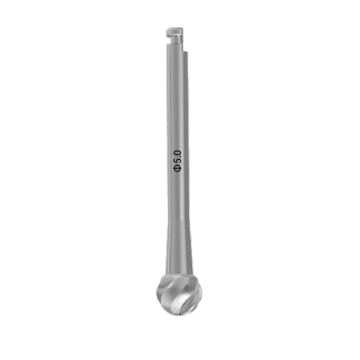
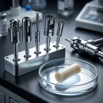
The hollow drill design minimizes bone loss and enables the collection of both autogenous bone powder and bone blocks for grafting.
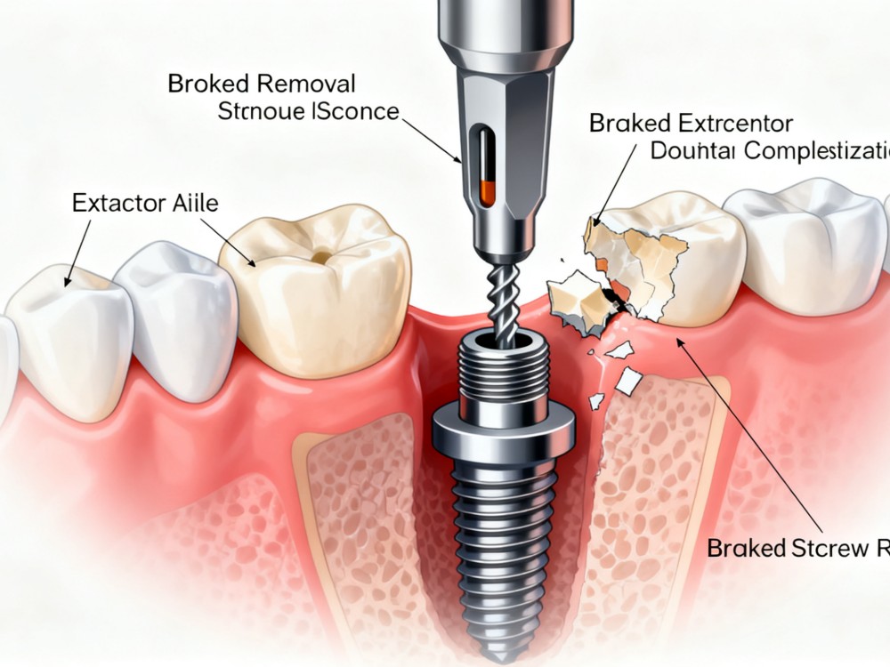
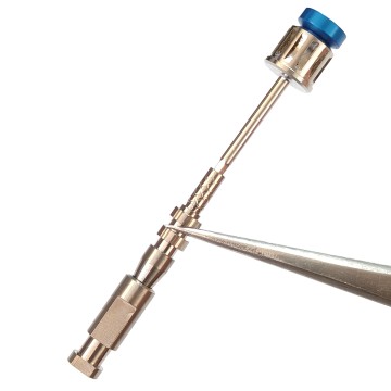
Comprehensive removal solutions for fractured abutment screws, central screws, or stripped screws, protecting the internal implant threads.
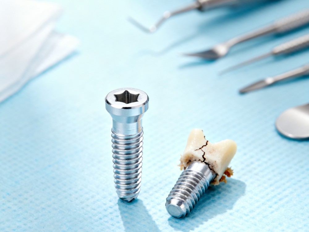
We offer advanced extraction solutions for removing both fractured/damaged implants and osteointegrated implants with minimal bone loss.
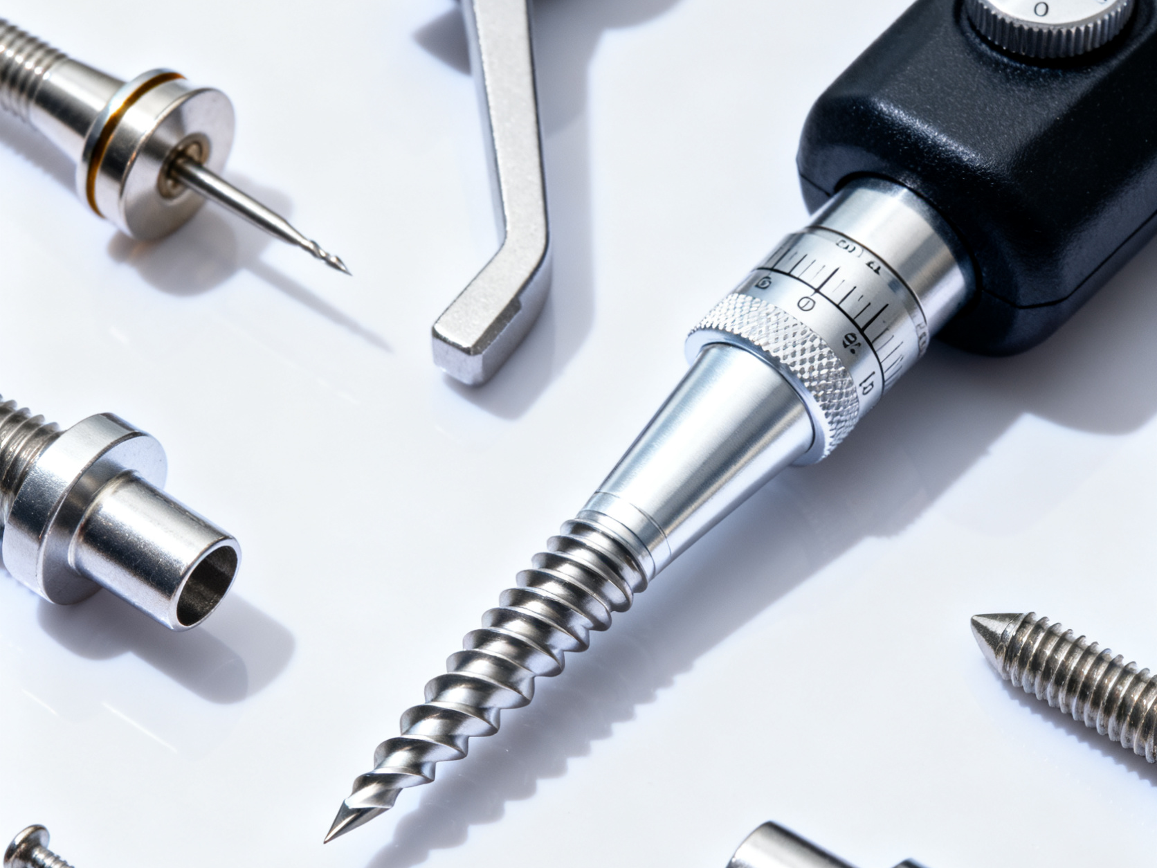
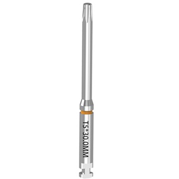
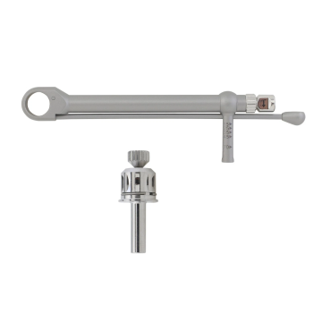
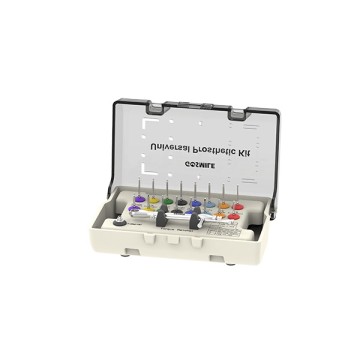
Our range includes drills, torque wrenches, and related accessories to support placement and analog replication of different implant systems.
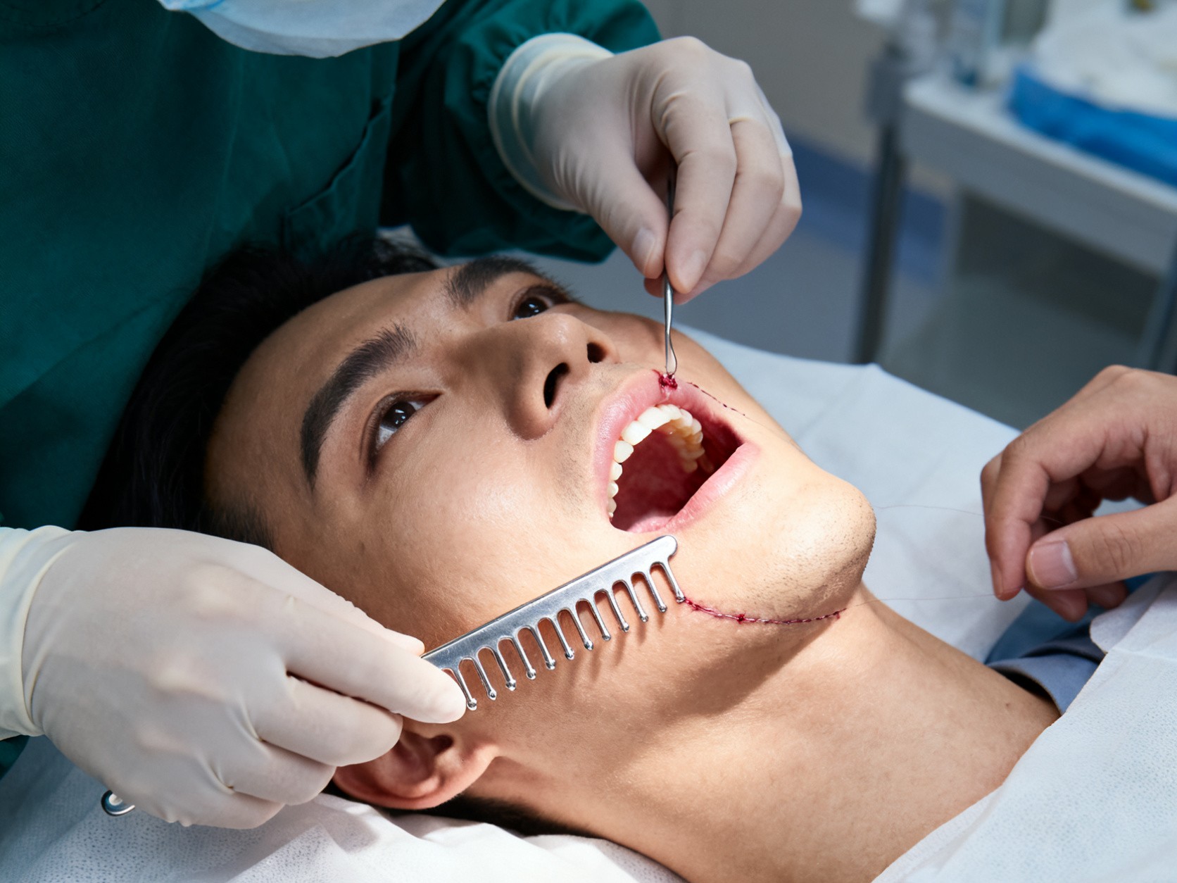
Our tension comb is designed for gingival soft tissue extension, providing adequate flap coverage over newly grafted bone sites.
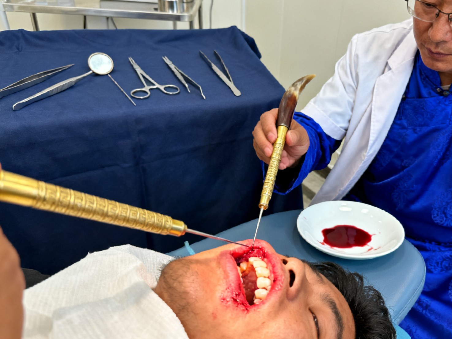
Our ultra-sharp triangular needles find applications in specialized therapeutic bloodletting and acupuncture procedures.
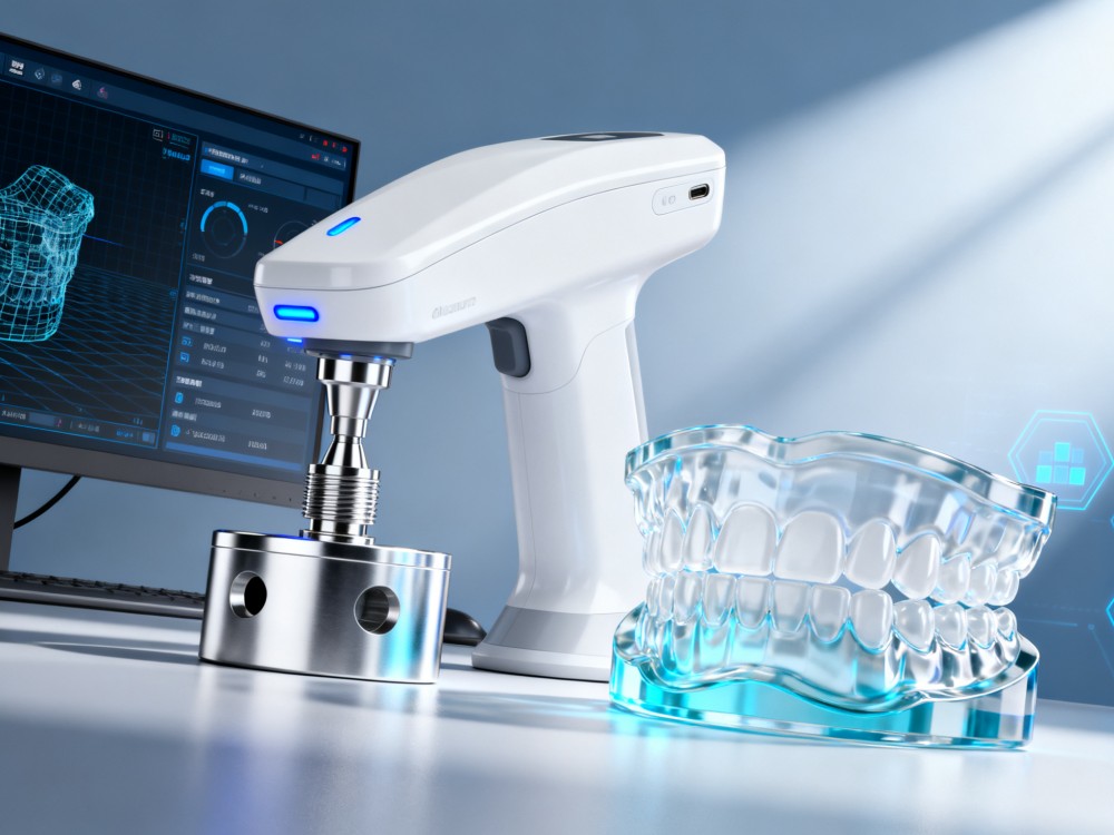
Customized scan bodies, scan abutments, and 3D analogs integrated with major CAD libraries to support a seamless digital workflow.
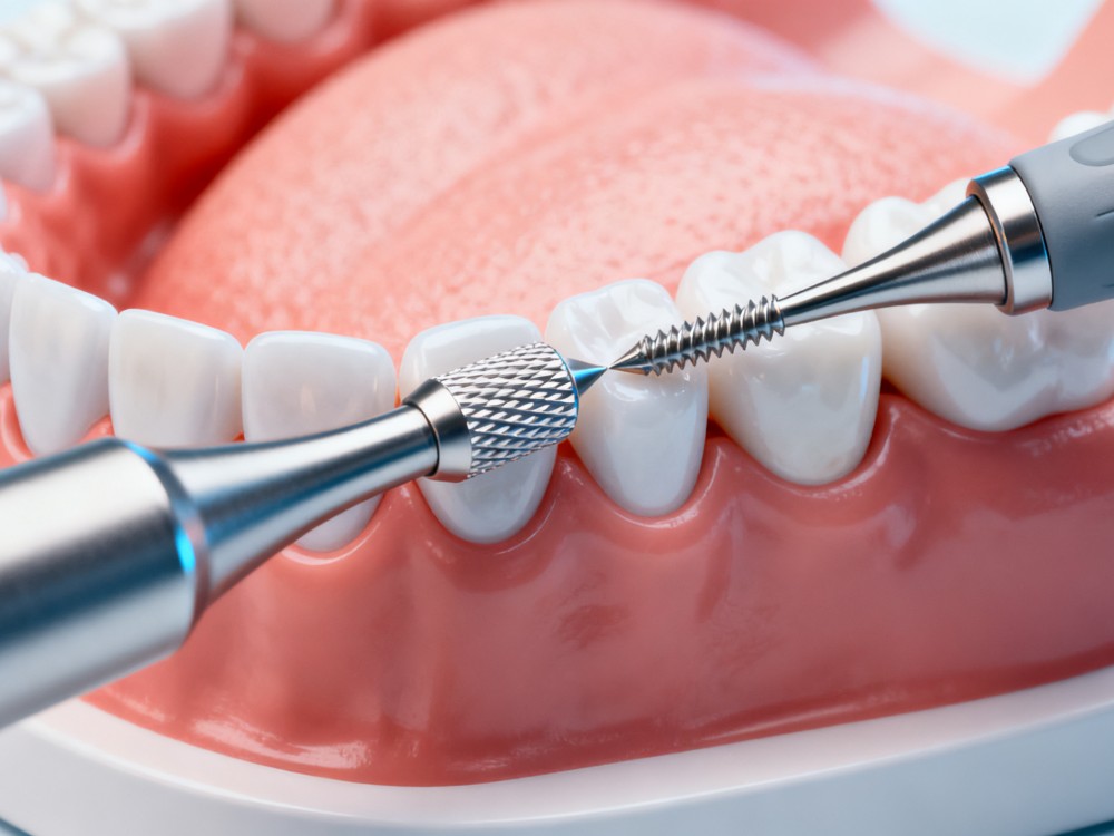
In combination with bone expansion drills, our tools facilitate controlled expansion and shaping of narrow alveolar ridges.
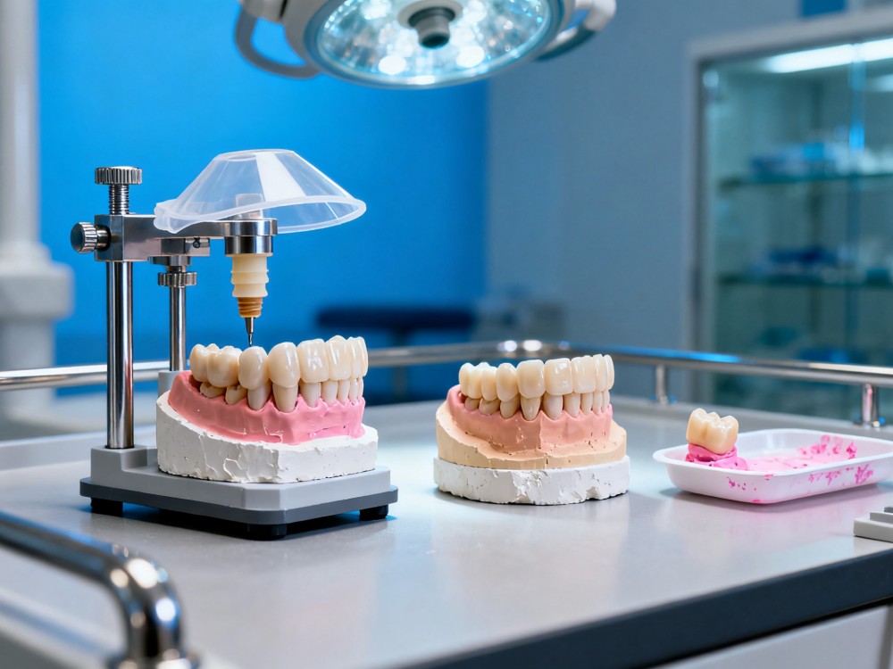
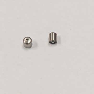
Our analogs, transfer posts, temporary abutments, and protective caps ensure complete accuracy from impression to model.

Providing high-fidelity training models and surgical kits to assist students and clinicians in simulated surgical practices.